Microalgae are actively used in the food industry, cosmetology, pharmaceutics, for cleaning water reservoirs, and as an alternative energy source. These raw materials are harmless for humans and the environment. At ITMO, ecologists study the promising applications of Chlorella vulgaris, one of the microalgae species.
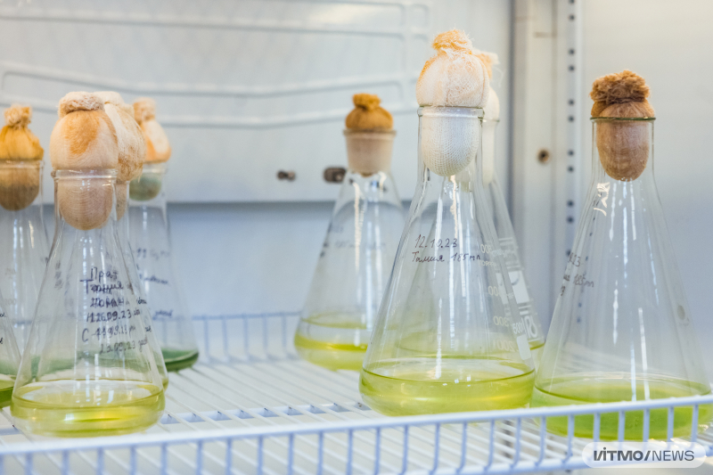
To cultivate microalgae on an industrial scale, it is necessary to determine the optimal conditions and medium for their cultivation. One of the most important indicators of growth and division of cells is their concentration. This marker reflects how many cells are present in one milliliter or gram of a substance, making it possible to compare different samples and identify the reasons for an increase or decrease in the number of cells. It is calculated manually using a method developed in the 20th century with the help of a Goryaev counting chamber – a marked microscope slide.
“This method is very time-consuming and effortful. In cases of high cell concentration, it takes up to 30 minutes to analyze a single sample, while usually you have to process over 10 of them! During this time, a researcher has to work with a microscope, processing each consecutive sector of the counting chamber, which often leads to lapses in concentration and errors. The only way to make this process quicker, cheaper, and more accurate is by automating it,” says Ivan Morshchinin, an author of the study and a PhD student at ITMO’s Faculty of Ecotechnologies.
Ivan Morshchinin. Photo courtesy of the researchers
There are currently dozens of similar tools on the market; however, the majority of them are neural networks that require large amounts of data for training, as well as experienced users and expensive equipment. Moreover, such AI tools can only detect the overall cell count in a sample, whereas their concentration still has to be calculated manually.
ITMO scientists have developed a method for automated gauging of cell concentration using images of samples. The software utilizes computer vision technologies and can be launched on a regular computer. All a user needs to do is upload microscope images of cells into a special folder and run the code. First, the photos are processed using algorithms: the color spectrum of the images is shifted toward green, then the images are converted to black-and-white and blurred. This is necessary to “clean” the images from solution impurities. Next, the Hough transform algorithm is “activated” to detect and separate circles in the image – since microalgae cells are round, the algorithm literally “outlines” them by their contours and separates them from each other. At the final stage, the identified cells are numbered, counted, and their concentration in the sample is determined with a special formula.
“Users can always manually check the quality of image processing. If the algorithm performed badly, it’s possible to change its hyperparameters – the microscope’s resolving power, the scale of the counting chamber, and others, and then run the code again. This is impossible to do with neural network-based tools. If just one feature of the images changes, an AI tool will malfunction – you’ll need to retrain it on new data. Our tool is adaptive and can work with images from any equipment and with any parameters,” explains Yulia Borisova, an author of the study and a junior researcher at the Research Center “Strong AI in Industry.”
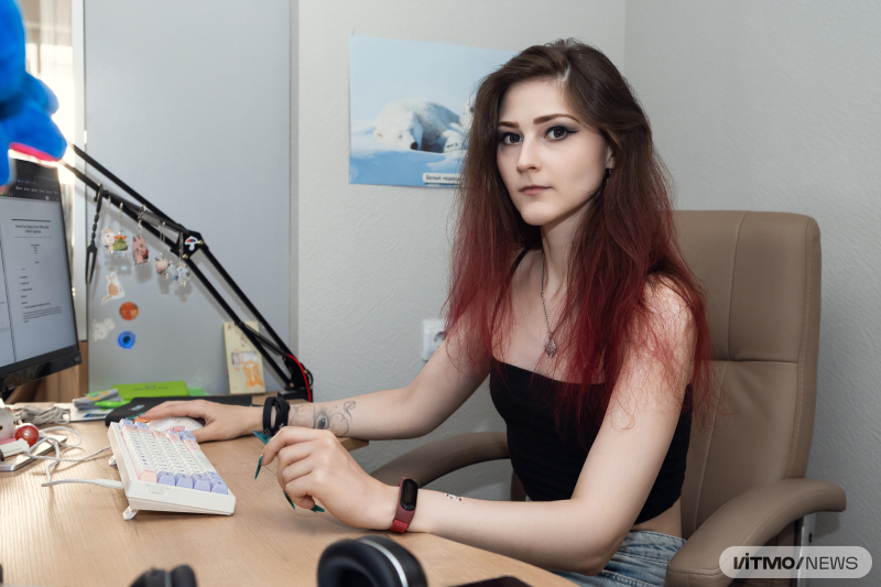
Yulia Borisova. Photo by Dmitry Grigoryev / ITMO NEWS
The new algorithm can process one “batch” of images in 30 seconds instead of the usual 30 minutes, which is 60 times faster than a human can. At the same time, as control tests showed, the quality of the obtained data remains the same and the likelihood of errors due to human factors decreases. Moreover, the algorithm identifies cells better than a popular pretrained neural network solution based on StarDist: 97% accuracy versus 92%.
According to Nelli Molodkina, an associate professor at ITMO’s Faculty of Ecotechnologies, who curated the study, the new method will significantly speed up the processing of Chlorella vulgaris microalgae. She states that AI and ML methods can improve the quality of ecological research, lower the error count, and expand the knowledge of objects studied thanks to new data.
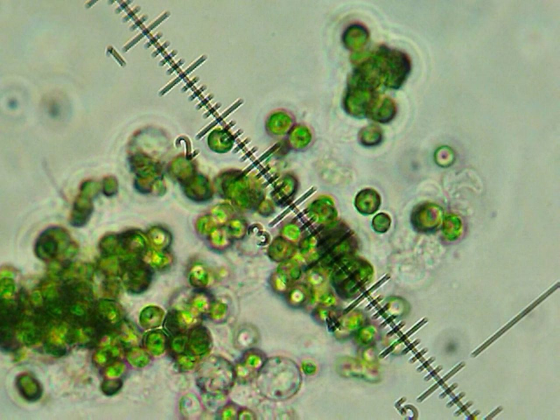
A microscope image of cells. Image courtesy of the researchers
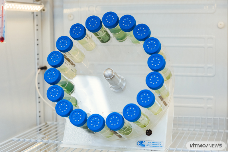
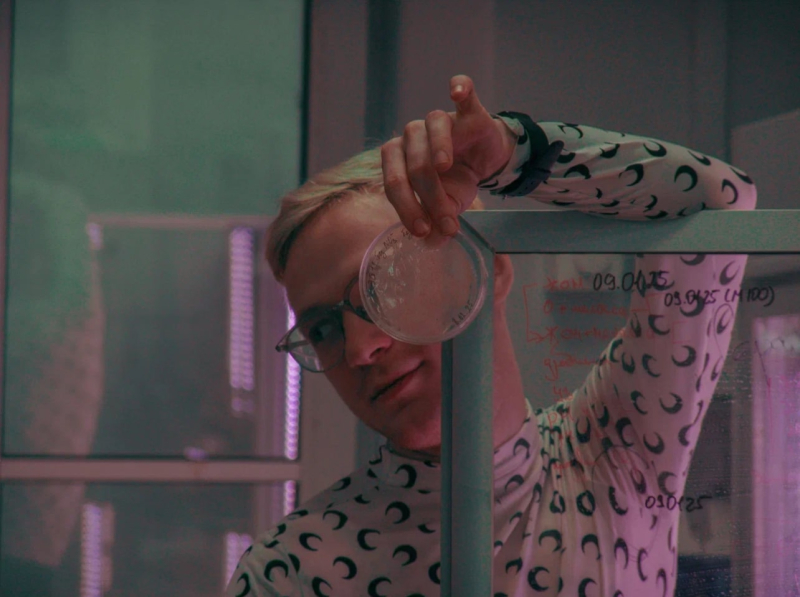
The setup for automatic calculation of microalgae cells concentration. Photo by Dmitry Grigoryev / ITMO NEWS
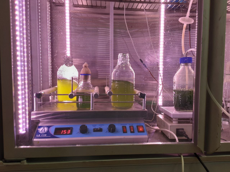
The setup for automatic calculation of microalgae cells concentration. Photo by Dmitry Grigoryev / ITMO NEWS
Next, the researchers are planning to turn their tool into a user-friendly application, as well as work with a different type of cells: for instance, blood cells, which are also round. In the future, this method could be used to process oval, spindle-shaped, flat, and other cells by replacing the Hough algorithm with a similar one used to work with a different type of data.
This study was the result of a collaboration between ITMO’s Faculty of Ecotechnologies and Artificial Intelligence Technologies Faculty, with students from both faculties – Yulia Borisova, Ivan Morshchinin, and Veronika Nazarova – joining in. The project was headed by associate professor of the Artificial Intelligence Technologies Faculty Nikolay Nikitin and associate professor of the Faculty of Ecotechnologies Nelli Molodkina.