With Raman spectroscopy, it is possible to accurately determine the chemical composition of various substances and their quantitative ratio in mixes. For this, the studied sample is excited with laser radiation at a specific wavelength; then, the light scattered by the sample molecules is registered. The resulting Raman spectra are unique for each compound, just like fingerprints.
However, the probability of Raman scattering is low, which is reflected in the method’s sensitivity: it is insufficient for detecting low concentrations of molecules, especially in complex media, such as biological fluids or petroleum products. This problem can be solved by using ensembles of nanoparticles that enhance the interaction of matter with light and, consequently, amplify the analytical signal. Most often, silver or gold nanoparticles are used for this purpose. Silver nanoparticles, although quite effective, are chemically active. This means that they can oxidize or interact with the substances being analyzed, distorting the results. The gold nanoparticles are more stable, but their effect is weaker; that’s why new approaches to signal amplification are being researched.
Physicists and chemists from ITMO University have developed composite materials, based on polymer microspheres coated with gold nanoparticles, that can be used to amplify the signal during Raman spectroscopy.
The scientists took two different approaches to creating these particles in order to see which would prove more efficient. Within the first approach, one that is widely used to produce nanomaterials, the microspheres were covered with a special polymer that acted as “glue” when the spheres were covered with gold nanoparticles.
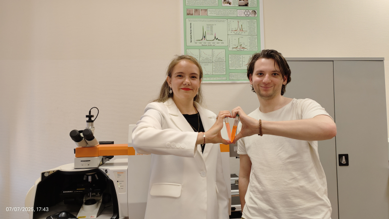
The paper's authors, Arina Pavlova and Anton Tkatch. Photo by Russian Science Foundation
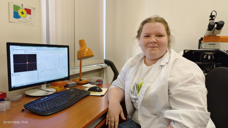
One of the paper's authors, Kseniia Maleeva, recording Raman spectra. Photo by Russian Science Foundation
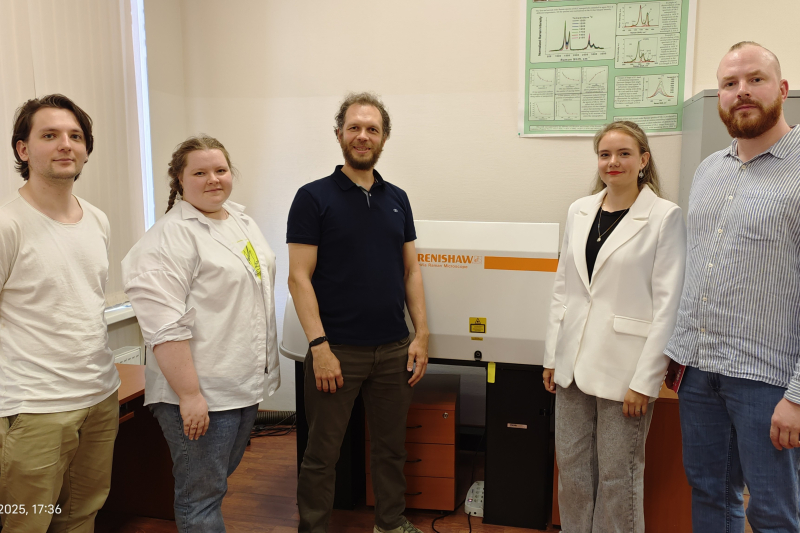
The paper's authors (left to right): Anton Tkatch, Kseniia Maleeva, Kirill Bogdanov, Anna Pavlova, and Evgeny Smirnov. Photo by Russian Science Foundation
For the second approach, the nanoparticles were stabilized with ascorbate, the salt of ascorbic acid, which meant that no additional polymer coating was required in the process – this made the preparation of signal-amplifying microspheres easier and cheaper.
The researchers analyzed the microstructure of the obtained spheres and the distribution of gold particles on their surface, while also confirming their experimental findings with modeling. Thus, ascorbate-stabilized nanoparticles formed dense clusters on the surface of the microspheres, which is crucial for enhancing the Raman scattering signal. Further investigation of the obtained spheres showed that the second method of applying nanoparticles ensures a denser coverage of the polymer sphere surface with gold and amplifies the signal by more than 14,000 times. For comparison, the microspheres obtained using the first, classical approach enhanced the signal only by 7,500 times.
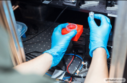
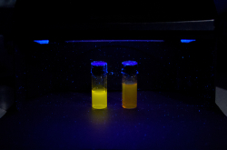
Additionally, the team tested the microspheres in real-world conditions, using them to analyze the quality of motor oil and detect dinitrophenol – a toxic compound used in the production of dyes and pesticides – in water. Thanks to the suggested “signal amplifiers,” the authors were able to use Raman spectroscopy to identify differences in the composition of fresh and used oil, which would be impossible to accomplish without the microspheres due to the strong fluorescence of the oil. What’s more, the new approach enabled the detection of dinitrophenol at concentrations 100 times lower than those accessible by conventional Raman spectroscopy.
“We acquired gold nanoparticles by switching sodium citrate, the conventional deoxidant, for potassium ascorbate, and noticed that such nanoparticles are metastable – with time, they aggregate and stick together. Next, we decided to try and precipitate the nanoparticles onto charged microspheres, which have long been studied by our physicist colleagues. As a result, we produced a great interdisciplinary study. I believe that the amplifying microspheres we suggest in our work are the perfect candidate for analyzing motor oil, determining its composition, and detecting impurities,” says Evgeny Smirnov, a professor at ITMO’s Infochemistry Scientific Center.
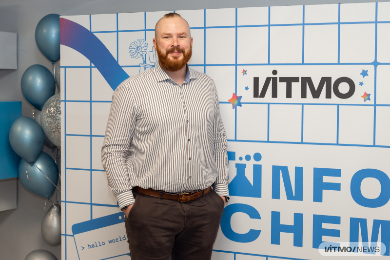
Evgeny Smirnov. Photo by Dmitry Grigoryev / ITMO NEWS
“The resulting composite structures significantly improve the method’s sensitivity, which means they can be applied when working with low-concentration samples or low scattering ability. Moreover, microspheres are much more convenient to apply in environmental monitoring or when assessing pharmaceuticals – for instance, detecting them in blood or analyzing their purity,” adds Kirill Bogdanov, the head of a lab within ITMO’s International Research and Educational Center for Physics of Nanostructures.
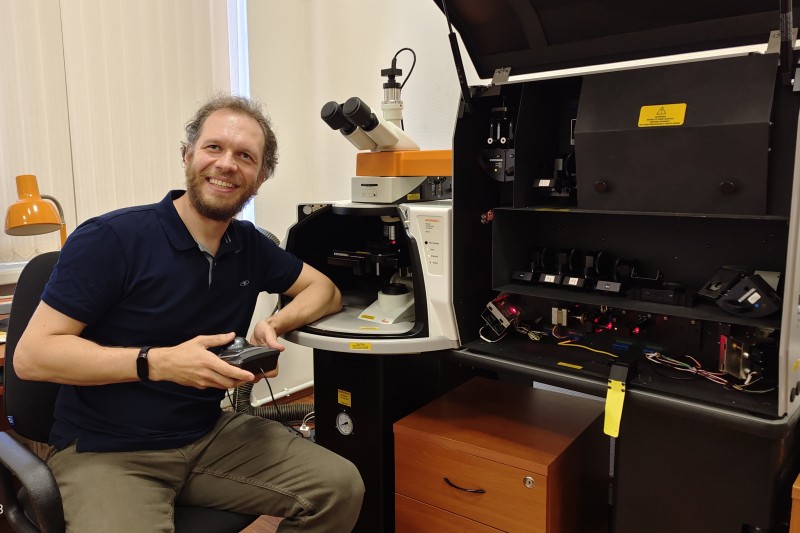
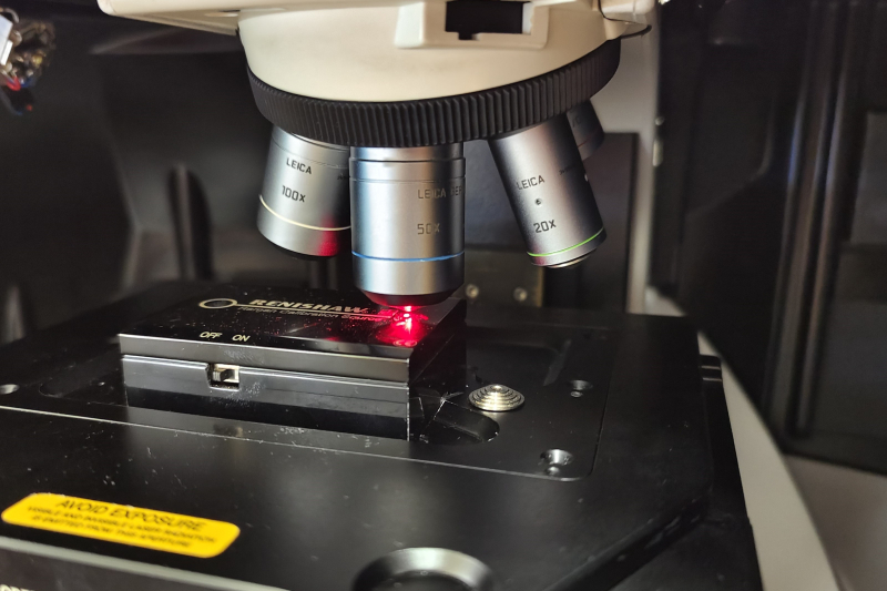
Kirill Bogdanov, the head of the project, by the inVia Renishaw Raman spectrometer. Photo by Russian Science Foundation
Next, the researchers are considering several vectors of study. They plan to add quantum dots (small semiconductor crystals) based on argon, indium, and sulfur to the composite materials. With these more complex structures, it will be possible to produce microresonators with a higher radiation intensity and quality. Such composites can be used to develop sensor platforms, as well as unique, unreproducible anti-forgery labels. Moreover, the researchers hypothesize that the acquired microspheres could be used in microfluid devices to detect pharmaceutical compounds in biological liquids.
This study was supported by the Russian Science Foundation grant No. 23-72-10010.
This article is created in collaboration with the Russian Science Foundation