Atomic force microscopes are used to study geometrical sizes of particles several nanometers in diameter, measure layers less than a nanometer thick, as well as analyze surfaces.
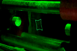
The microscope works through “palpation” of the surface with a probe that is located on the end of a long elastic rod – the dark narrow stripe seen in the picture above. The microscope is called atomic because it functions thanks to the force interaction of atoms.
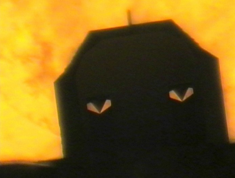
The photograph seen above was produced by a camera installed inside the microscope. The yellow background is formed by a research sample. This particular shade of yellow is produced by a lamp working at low capacity in order to conserve its resources and avoid overexposure.
The picture was taken during the setting-up of equipment. Before researchers start using it, they need to direct a secondary laser’s beam onto the top of the probe. It can be adjusted via a signal from a photodetector or by looking through a secondary microscope. Moreover, with the help of a secondary microscope you can see which part of the sample is located under the probe. Therefore, you can choose a specific part of the sample to study. That’s what scientists were doing when they noticed the probe’s resemblance to a scary mask and decided to take a picture.
The microscope is used by ITMO University’s International Research and Education Center for Physics of Nanostructures to measure the thickness of metal films, nanoparticle layers, and nanoparticles about 3-10 nm in diameter. With its help, the scientists determine the diameter of carbon dots that are researched as part of the “Light-Emitting Carbon Dots” grant. The properties of carbon dots can be modified via extra processing after the synthesis. If their size changes, then the processing went well.