MR image of the whole body is sometimes required for the body systems studies, as well as for tracking the effect of various drugs. Most often preclinical studies are conducted on animals, for example, on mice. Despite the small size, obtaining high-quality image of the whole mouse is not as easy as it seems.
Usually, researchers use standard volume MR coil to emit a radiofrequency signal and individual small coils to receive a signal from the area of interest. Therefore, to get images of the whole body, one need to either combine images from local coils, or use the standard volume coil for both transmission and reception. In the first case, the image obtaining procedure becomes more complicated, while in the second one, the quality of the image is so poor that it becomes difficult to distinguish small important details.

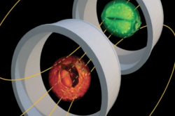
To solve this problem, researchers from ITMO University developed a new type of special MR coil. A number of its design advantages makes it possible to get a high-quality image of the whole mouse. First of all, the coil has an optimal size and field of view for mouse imaging. A metastructure with a distributed capacity at the base of the coil provides an ability to choose the size of the resonator tuning it to the dimensions of the mouse in order to avoid extra noise reception. In this case, the radiofrequency magnetic field intensity in the area of interest is much larger than the one produced by the standard volume coil. It provides a uniform spatial distribution of the received signal. This leads to a high sensitivity of the coil in the entire field of view and improves the image quality.
"Standard coils are tuned to the desired frequency with variablenon-magnetic capacitors. Since their usage introduces high internal losses, the signal-to-noise (SNR) ratio is reduced. SNR is one of the main parameters that determine the image quality in MRI. We do not need any capacitors for our coil as it is self-resonant and may be tuned by changing its geometric parameters. The coil design allows us to optimize its operation, increase the sensitivity and image quality. In addition, the cost of materials used is low, and the manufacturing technology may be adapted for different studies," says Anna Hurshkainen, a PhD student at ITMO University, researcher in the Laboratory of Nanophotonics and Metamaterials.
According to researchers, the work started with numerical modeling. This helped to optimize the geometry of the coil, as well as to choose materials. After that, the researchers made a coil prototype and conducted experiments on the phantom and mice.

"We measured the signal-to-noise ratio in different parts of the image at different distances between the object and the coil. The obtained results were compared with the simulation and with similar numbers for the standard volume coil. As a result, large distance from the object to the coil reduced difference almost to zero, while at a minimum distance, the signal-to-noise ratio of our coil was better by 60%. We also managed to achieve a threefold improvement in quality in the optimal distance," adds Mikhail Zubkov, researcher at the Laboratory of Nanophotonics and Metamaterials of ITMO University.
Currently, researchers plan to continue working on a variety of coils for various preclinical studies.
Reference: Small‐animal, whole‐body imaging with metamaterial‐inspired RF coil. Mikhail Zubkov, Anna A. Hurshkainen et al. NMR in Biomedicine, 26 June 2018


