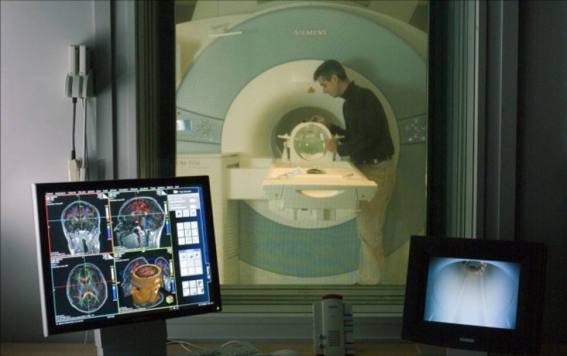
Magnetic resonance imaging (MRI) is a technique for obtaining tomographic medical images for the examination of internal organs and tissues using the magnetic nuclear resonance phenomenon. The procedure is based on the excitation of nuclei with a special combination of electromagnetic waves in a high-intensity, permanent magnetic field. Using MRI allows medical professionals to diagnose tumors at an early stage, examine severe pathologies of the cardiovascular and respiratory systems, and perform other crucial imaging.
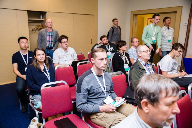
But medicine is not the only field that benefits from MRI. Experts from Radboud University Nijmegen, Western Sydney University, and Aix-Marseille University, as well as other prominent research institutions, weighed in on these different applications of MRI, as well as work to improve the technology, at the BioMETANANO section of the fourth METANANO conference recently held in St. Petersburg.
For one, Professor Arend Heerschap, of Radboud University Nijmegen Medical Center, the Netherlands, spoke about his and his colleagues’ research involving MRI and nanoparticles. The scientists work with small particles of iron oxide, packaging these in dextran, a high molecular weight carbohydrate commonly used in medicine to lower blood viscosity. The altered iron oxide particles are then introduced into a patient’s blood, facilitating the detection of various cells in the patient’s lymph nodes using MRI technology.

Professor William Price from Western Sydney University, Australia, gave a presentation on the materials analysis method nuclear magnetic resonance (NMR) diffusometry. Diffusion is the process wherein the molecules of one substance move from an area of high concentration (where there are many molecules) to an area of low concentration (where there are fewer molecules) of another until the system has reached equilibrium (molecules evenly spread). According to Prof. Price, putting this process under examination can result in valuable new information on the size of the molecules involved, as well as their environment and movement patterns. All this is very important for the field of bionanotechnologies.
“Depending on their professional background, specialists tend to view MRI from different angles. Clinicians use the technology for finding tumors in the brain; chemists – for identifying compounds, to see what they ended up with; biochemistry specialists – for understanding the molecular conformations. The study of diffusion can open up a whole new world of new activities and fields, from neurobiology, electrochemistry to crop farming and many others,” says Prof. William Price.

In his turn, David Bendahan from the Center for Magnetic Resonance in Biology and Medicine of Aix-Marseille University, France, focused on how scientific literature sees the future of magnetic resonance imaging. The expert’s own forecast is that the technology will significantly ramp up its capacity. The existing devices operate on the capacity of 1.5 or 3 T, or Tesla, a magnetic induction and magnetic flux density measuring unit named after the Serbian-American scientist Nikola Tesla. The Tesla value depends on a magnet used: the higher it is, the better is the quality of a resulting image. According to the researcher, the future MRI equipment will work with the capacity of 7 T. This change is expected to lead to a number of significant improvements: the devices will also be able to record an image faster, producing it in better resolution and contrast.


