In order to fully provide tissue and organs with all necessary substances and oxygen, we need not only a branched network of arteries that conducts this delivery to the heart, but also its correct operation. Coronary arteries supply the tissue of the heart itself. Pathological changes in these arteries lead to a worsening of the blood circulation system of the heart. A consequence of such changes and disorders may cause various cardiovascular diseases such as ischemia and acute coronary syndrome. These diseases and concomitant damage to the heart muscle (the myocardium) are usually caused by a pathological narrowing of the coronary vessels.
For a long time, only surgical intervention by chest dissection could be used to treat narrowed coronary vessels. However, in recent decades, less traumatic methods have emerged and begun to be widely used, allowing access to the narrowed vessel by inserting a catheter into an artery. One of them is coronary stenting of blood vessels.

Coronary artery stenting is one of the safest and most effective procedures for restoring the normal lumen of arteries that have atherosclerotic changes. The stent is a reticular cylindrical metal frame which unfolds in the occluded part of the artery and prevents it from narrowing again. After installation, the stent is naturally covered by the inner lining of the vessel.
However, this procedure is not always successful. In some cases, restenosis develops - a complication that is characterized by excessive tissue overgrowth and narrowing of the artery lumen again. Nowadays, the causes of restenosis are being studied. There are researchers that aim to optimize the configuration of the stent and the treatment procedure to reduce the number of complications. But how can computer modeling be helpful in this situation?
One way to determine the causes of restenosis is to conduct experiments using cell cultures and model animals. This kind of studies, for example, are is carried out at the University of Sheffield. However, computer modeling can help in understanding the causes of restenosis as well. Moreover, it can be used to design a more effective stent construction that will reduce the amount of complications.

The same research is being carried out by specialists of the eScience Research Institute at ITMO University, who in collaboration with their colleagues from Amsterdam University in the Netherlands and University of Sheffield in the UK have been working over the last few years to build a model of undesirable growth of tissue inside the vessel after the stent (expanding frame) was installed in it.
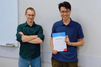
Pavel Zun explains why this model can help to understand why in some cases the tissues grow too intensively through the stent and the vessel re-narrows, causing restenosis. By reproducing this process, scientists learned to model the cellular growth of the arterial wall, the physical interactions that arise from the fact that the stent is stretching the vessel, and also how the medical substances from the stent penetrate deep into the tissues.
Within the framework of the project, Pavel Zun spent the last month at the University of Sheffield, where he worked on further improvement of the model validation of the results obtained in the course of computer modeling.
The process of designing the model is as follows: first, scientists conduct experiments. For example, they take cells and plant them in Petri dishes, and then they watch how these cells behave under the influence of currents, how they migrate, and so on. Based on the data obtained from a large number of experiments, a hypothesis is constructed about what mechanisms influence the behavior of cells. And, in turn, based on this hypothesis, a model is constructed: we establish certain rules for the behavior of cells in this model, depending on what we want to check. After that, we check how much the behavior of the cells in the model with a certain set of rules corresponds to what is happening in reality, - says Pavel Zun. - Thus, my work in Sheffield consisted of finding experimental data, as well as of cooperation with biologists. After all, we can build a model, and they can put relevant experiments in vitro, on cell cultures, or on animals, in vivo. In the future, these experiments can be used to validate our models.
One of the main factors that stop tissue growth in the vessel is the restoration of the cells of the inner lining of the vessel - the endothelium. Thus, in order to describe the processes occurring in the vessel, it is necessary to know how the endothelial cells migrate after the stent has been installed and how the healing process takes place. This can be studied by putting these cells into an experimental setup that mimics the stented part of the vessel and tracks how they will move there under the influence of currents. By studying these processes, the trajectories of the movement of cells can be obtained and then used by researchers in the construction of computer models. As Pavel Zun points out, in recent years scientists have managed to achieve quite a good accuracy of the model.

For example, with the help of modeling we see that between the ribs imitating the stent elements placed on the surface of the installation, there are vortices of the fluid flowing through the installation. This affects the trajectory of cell migration, and we can speculate why this happens, - explains the researcher. - Accordingly, based on the simulation, we can offer a new design of the experiment to test this assumption. If this assumption is correct, this will affect the stent design, which in the future will reduce the risk of complications after surgery.
The risk of complications after the installation of the first stents, which began to be used in the 90s of last century, reached 20%. Modern devices helped to reduce this figure to five cases out of a hundred. However, considering that stenting of the coronary vessels is one of the most common operations in modern medical practice, further improvement of the stent design and reduction of the risk of complications is still a relevant objective for researchers, says Pavel Zun.

Today, scientists continue their work on improving the model. In the near future, researchers are going to test another hypothesis, according to which the acceleration of the healing of the artery after stenting can be affected by the coating of the stent with a special substance that suppresses Rho-associated protein kinase in cells (ROCK), responsible for the reaction of cells to the local direction of the blood flow. This mechanism will also be implemented in the model and, based on the results of the work, will show how much it will match the experimental studies.
Potential use
The construction of tissue growth model inside the vessel after stent installation is a part of the work that scientists in Amsterdam and Saint Petersburg have been conducting in the last few years. In an earlier study published in the journal Philosophical Transactions A, they presented the concept of a "virtual artery", showing that this multiscale computer model would combine several submodels that describe the parts of the cardiovascular system in different approaches. The developers believe that a detailed simulation of the human artery will allow a deeper study of vascular disease and will create an alternative to animal testing.
A fully complete virtual artery, according to scientists, will allow studying a wider range of cardiovascular diseases, combining several models of different levels. At the first level, the developers reproduce the bloodstream throughout the body. As they go deeper, they enter a three-dimensional section of the blood vessel. At the lowest level, interactions between cells within the arterial wall are observed. Thus, step by step, scientists approach more accurate reproduction of complex natural processes.
At the current moment, scientists of ITMO University are proceeding with their collaboration with the scientific group of the University of Amsterdam, as well as with the specialists of the University of Sheffield. In addition, they recently established new collaboration with researchers from the Polytechnic University of Milan, who are also involved in the modeling of currents in stented vessels - says Pavel Zun.

According to him, the construction of computer models for medicine is one of the most interesting and promising tasks today. A large number of new projects have appeared recently in this field. And there are prospective graduates and post-graduate students who can apply their powers to such projects.
It is very interesting to be engaged in modeling precisely in the field of biochemistry, medicine, because modeling in this field is not yet developed as much as, for example, in physics or abiochemistry. These are very complex systems, so there is still an unexplored field for research. Yes, already in the 90's there were attempts to model systems in this area, but then, of course, there was not enough computational power to make complex models, – says Pavel Zun. – But the most important resource which this area lacks is people. Not many graduate and post graduate students are engaged in these programs. Meanwhile, there are a lot of projects on this subject today, so the demand for qualified specialists is large.


