Microparticles that emit light under the influence of electromagnetic radiation find their applications in biosensors and optics. Thanks to their fluorescence, it is possible to monitor targeted drug-delivery within the body in real-time, as well as to detect tumors by registering the fluorescence of antigen protein clusters around cancer cells.
Instead of traditional organic pigments, quantum dots – small crystals of materials capable of conducting electricity when influenced by temperature or light, can be used to achieve brighter fluorescence. Among such materials are silicon, cadmium, selenium, and other semiconductors. Under UV radiation, electrons in quantum dots get excited to the state with higher energy that is later released as photons. This effect is what creates fluorescence.
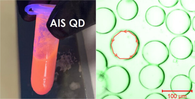
Scientists from ITMO have suggested a method to produce low-toxicity fluorescent polymer microparticles with quantum dots; the method is based on forming isolated substance droplets in a flow of microscopic volumes of liquid. The study was conducted in collaboration with St. Petersburg Academic University and the Institute for Analytical Instrumentation of the RAS. Kamilla Kurasova, a graduate of the Master’s program Physics and Technology of Nanostructures, became the first author of the corresponding paper.
In lab conditions, the research team has designed a system of intersecting channels that works with smaller volumes of liquids and makes it possible to control them. The device creates polymer microspheres with quantum dots inside – nanocrystals based on silver, indium, and sulfur. The size of the spheres is comparable to the thickness of a human hair, which is just 55-95 micrometers.
In order to stimulate the fluorescence of microparticles, the quantum dots were excited with a laser; then, the researchers observed the fluorescence intensity and its fade-out time. With the new approach, it is possible to produce brightly glowing particles that are resistant to lasers. This means that particles can be repeatedly excited during long experiments while maintaining their fluorescence. At the same time, the duration of quantum dots’ fluorescence decreased to 3.5 nanoseconds, which is tens of times shorter than the fluorescence in a thin polymer film. This short fluorescence interval will enhance the accuracy of monitoring processes in the human body, as the shorter the time a quantum dot remains in an excited state, the less it is influenced by the surrounding environment.
“Our suggested method of microparticle production may be widely applied in biomedicine. Conventionally, quantum dots are made from toxic heavy metals and diluted in aggressive environments – in our study, we used low-toxicity quantum dots. Thanks to this, it will be possible to apply our microspheres with quantum dots as tools to determine blood flow, intravascular visualization, calibrate equipment, deliver treatments and enzymes in the body, and more,” explains Anton Starvoytov, an associate professor, senior researcher at the laboratory Surface Photophysics of ITMO’s International Research and Educational Center for Physics of Nanostructures.

Anton Starovoytov. Photo by Dmitry Grigoryev / ITMO.NEWS
The research team continues their analysis of the microparticles. They are being introduced into lab animals, where the fluorescence of quantum dots is triggered by laser and diodes. At this stage, it’s crucial to ensure the low toxicity of the microparticles. If this is proven, researchers can discuss the possibility of proceeding to tests on humans.
The results of the study were supported by the Russian Science Foundation grant No. 22-72-10057.


