The first one in Europe, the second one worldwide
Evgeniy, tell us, why was this facility assembled in France?
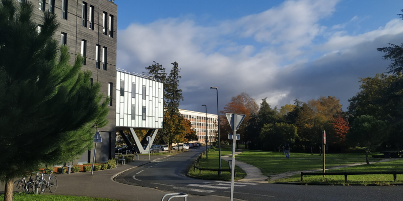
University of Bordeaux scientists have been working in this field for several years now. They had the necessary laboratory equipment, parts of the mechanism and materials, but no specialist who would assemble it. Due to the fact that my thesis advisor, Olga Smolyanskaya, head of ITMO University’s Femtomedicine Laboratory, has been cooperating with the University of Bordeaux for three years, and has several shared projects, they offered a position for the staff of our laboratory. That’s how I got the internship.
What is the main working principle of the system?
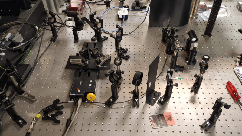
It’s an optical facility based on a femtosecond laser. The wavelength of terahertz radiation is quite big, so it’s hard to get images in the terahertz range with quality that would allow one to see small features of a studied object. In order to achieve high quality, you need to focus the laser beam, so that the diameter of a spot in focus would be as small as possible. As there are no facilities of that kind in Europe yet, and there’s only one of them in Japan, I used the LTEM (Laser Terahertz Emission Microscope) method as proposed by Prof. M. Tonouchi.
So there are no similar systems in Russia at the moment?
As far as I know, there are no facilities based on Tonouchi’s principle. However, similar research is going on at the laboratory of Prokhorov General Physics Institute of the Russian Academy of Sciences, under the supervision of Kirill Zaitsev, but they use another method. I’m sure that many researchers in the terahertz field would like to have pictures of high quality, therefore they aspire to create a similar technology.
It’s a new topic that started to develop after M. Tonouchi, a Japanese scientist, published papers on it. I think a system of that kind can be assembled at ITMO University, too. We cooperate a lot with Valerii Trukhin, a research associate at ITMO University’s Faculty of Photonics and Optical Information, who is working on a terahertz microscope. I’m sure that by putting together our experience and expertise we could get a good result.
In France, were you assembling it alone or as part of a research team?
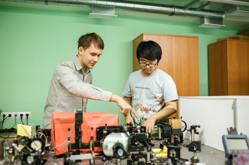
My thesis advisor was supervising the assembling, and Frederic Fauqet, one of the laboratory’s employees, was helping me with the arrangement, for example, with purchasing the equipment and parts of the mechanism, but the rest of the work was on me.
Interrupted research
Your internship ended one and a half months earlier than it was supposed to due to the pandemic. Tell us, at what point have you stopped?
The job is 80-85% done. We managed to receive a terahertz signal and to assemble the necessary components of the system. We only have to solve a problem related to disperse distortion in the optical fibre, a part of the detector that is responsible for getting a probe beam to the terahertz signal detection area. I hope I’ll be able to continue the work when we are able to go back to the laboratory again. We’ve discussed it with the head of the project, Jean-Paul Guillet.
Do you somehow continue the work remotely?
I use this break to write various texts on the project: descriptions of the work process and reports. I do what I can remotely, but mostly it’s a practical task that requires me being in the laboratory.
Is it your first research abroad?
Yes, and it was highly beneficial. I’ve learnt a lot both as a person and as a researcher. I found out what it’s like to live and to be a scientist in France. There were certain obstacles, but we overcame them. Now I have a lot of experience and I feel much more confident in assembling complex optical systems.
What kind of research are you planning to conduct in the future?
In my thesis, I want to prove the possibility of getting pictures of cancer cells in the terahertz frequency range with a high spatial and longitudinal resolution. Physical and mathematical models of the interaction of THz radiation with biological objects will be proposed, and new methods of pulsed terahertz spectroscopy and microscopy with enhanced technical characteristics will be developed.
Our laboratory cooperated with N.N. Petrov National Medical Research Center of Oncology and Almazov National Medical Research Centre. They share biomedical objects with me and consult me on the theoretical basis of detected effects, as well as on how the research can be applied in clinics. Results that I will get may be used in order to create a new method for monitoring pathological changes in body tissues.
