Recently, Dr. Dunaev joined the ITMO Fellowship program and, on the invitation of ITMO’s Scientific and Educational Laboratory “Machine Vision,” conducted a special course for the university’s students.
You’ve been in biomedical photonics for over 20 years. What is this field and what prospects does it open up for us?
As a scientific discipline, biomedical photonics studies the interactions between photonics and biological objects such as blood vessels, tissue, and organs. These days, almost everyone has a fitness band or a smartwatch to measure their heartbeat; in the days of the pandemic, everybody got themselves a pulse oximeter to measure their blood oxygen levels. These are just the simplest examples of wearable biomedical photonics devices.
All in all, our research allows us to track a lot of health metrics in the human body and diagnose various illnesses. For instance, we can use LEDs or lasers to assess tissue saturation, blood content, and perfusion (the passage of blood through tissue) or gauge the melanin index, which is responsible for protecting our skin from harmful UV radiation.
All in all, I have, indeed, been involved in biomedical photonics research for over 20 years. I’ve defended my PhD and DSc theses in this field. In 2010, we established an eponymous R&D center at Orel State University. There, we try to integrate biomedical photonics-based engineering technologies into the operations of various medical organizations.
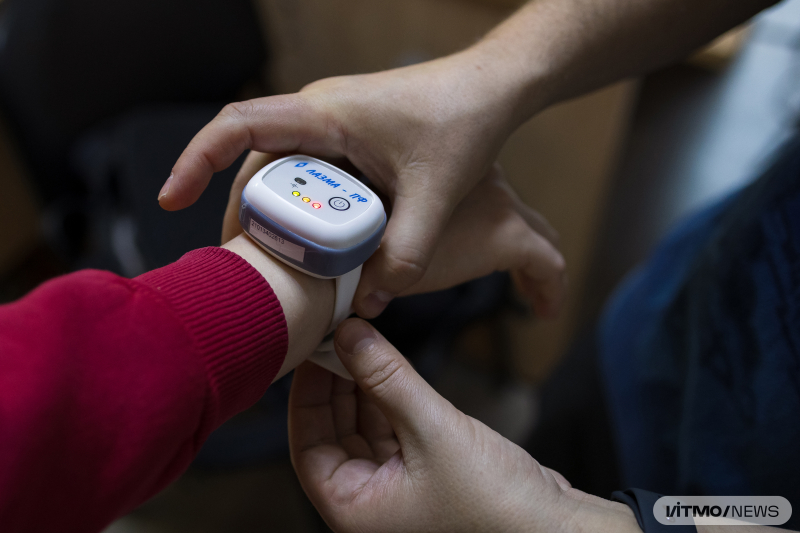
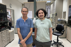
Andrey Dunaev and his students measure their peripheral blood flow with the help of a portable microcirculationtester. Photo by Dmitry Grigoryev / ITMO.NEWS
Andrey Dunaev and his students measure their peripheral blood flow with the help of a portable microcirculationtester. Photo by Dmitry Grigoryev / ITMO.NEWS
Andrey Dunaev and his students measure their peripheral blood flow with the help of a portable microcirculationtester. Photo by Dmitry Grigoryev / ITMO.NEWS
What are these technologies?
Recently, we finalized a project on post-COVID rehabilitation, which we’d worked on together with the Ministry of Healthcare’s National Medical Research Center for Therapy and Preventive Medicine. Since the pandemic, people have had complaints about fatigue and memory issues. In order to assess the changes in microcirculation of blood and tissue perfusion in the forehead area, we asked patients to complete a few cognitive tests – for instance, to find objects in a specific order in a Schulte table (named after its creator, the German psychiatrist Walter Schulte). Meanwhile, us researchers would measure the subject’s peripheral blood circulation.
Usually, these measurements are taken around the fingers or the forearm, but we suggested using the forehead area instead. The study showed that patients with post-COVID complications had a lower level of skin blood flow in their head. This may be connected, among other things, to the cognitive effects of post-COVID syndrome, such as issues with memory, concentration, and so on.
You’ve mentioned diagnostics. There are many well-known and well-tested methods out there. What advantages does biomedical photonics offer?
The most important one is its non-invasive nature. Unlike histology or endoscopy, these methods are safer and less traumatic. They can be used to detect pathological processes at earlier stages and to study objects under a millimeter in size, which cannot be detected via ultrasound, CT, or MRI. Besides, biomedical photonics methods are safe for patients who are pregnant or have medical limitations.
Another advantage is that these methods have a wide range of applications. Together with the Ott Research Institute of Obstetrics and Gynecology, we’ve studied diabetes-related complications during pregnancy, which are associated with metabolic changes that cannot be easily traced. Using portable analyzers, we’ve collected data on the patients’ peripheral blood flow and prescribed the appropriate treatment that would minimize such complications.
We’re also using wearable sensors to monitor sleep quality and to study the how and why of insomnia. These days, electroencephalography is used for this purpose. This means the patient needs to wear a headcap with a massive mane of wires and has to fall asleep so that their brain activity can be analyzed. We’re trying to replace this uncomfortable method with portable sensors that monitor blood perfusion and transmit data over Bluetooth. As of now, we’ve recorded data from 15 volunteers and are processing it.
You can also use the methods of biomedical photonics for minimally invasive surgery. We were once contacted by a surgeon who does transdermal needle liver biopsy. With the help of ultrasound, a special needle is inserted into the organ in order to take a biopsy sample. Through cytological and histological tests, it’s possible to assess the nature of a liver tumor. But the issue is that the precision of the needle’s positioning depends on the surgeon’s level of experience. The error rate may reach up to 25-30% – and you can’t take a second sample on the same occasion. To make sure the surgeon can get to the tumor, we’ve enhanced the needle with biophotonics tech that allows the user to analyze various parameters of the tissue (saturation, fluorescence amplitudes of endogenous fluorophores) and more accurately pinpoint the area of the tumor. As a result, we’ve improved the accuracy of the procedure to 90%.
All in all, we adopt a multimodal approach, combining several methods to obtain more reliable and comprehensive data about the disease. When medical specialists tell us about their challenges with diagnostics, we first study the object of research – the microcirculatory systems of the human body – and then select the appropriate technologies.
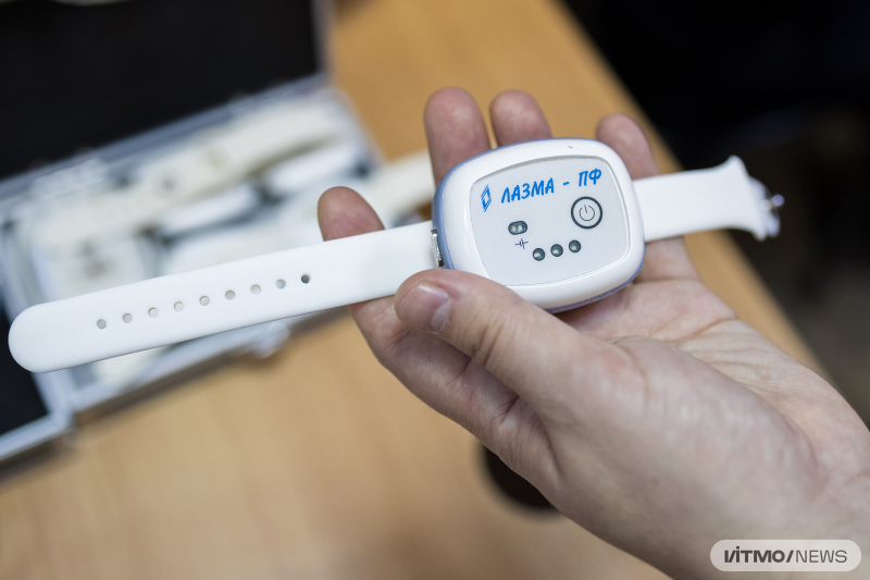
ITMO students measure their peripheral blood flow with the help of a portable tester. Photo by Dmitry Grigoryev / ITMO.NEWS
ITMO students measure their peripheral blood flow with the help of a portable tester. Photo by Dmitry Grigoryev / ITMO.NEWS
The threshold for entry into biomedical photonics seems very high. What does one need to know if they want to work in this field?
It’s a wholly interdisciplinary science. I usually say that we have one foot in biomedicine and the other in engineering. You can’t stay on just one side. Additionally, you need to understand both medical and "regular" physics. Therefore, the ideal option is to complete a Bachelor’s degree in medical technologies or physics and then a Master’s in biology or biophysics – or vice versa. In other cases, it’s better to take some professional development courses if you’re truly interested in this field.
How do you predict the field will develop in the future? What new opportunities or breakthroughs can we expect?
The main drawback of biomedical photonics is that light cannot penetrate very far into tissue or organs – only a few millimeters. In other words, the depth of biomedical diagnostics is quite limited. To solve this problem, scientists are conducting experiments with various reagents that make biological tissue more transparent.
For example, American researchers have already managed to make the abdomen of a living mouse transparent in order to observe its organs. In Russia, the method of tissue clearing was devised and is being developed by Valery Tuchin, a prominent scientist and one of the founders of biophotonics. He is currently the head of the Department of Optics and Biophotonics at Saratov State University and a corresponding member of the Russian Academy of Sciences. He suggested applying reagents (such as glycerin) to the skin to displace the water from it and create a homogeneous medium for tissue visualization. For his contribution to science, Tuchin was awarded the national Challenge prize last year in the Scientist of the Year nomination. Of course, with this technology, we won’t become "invisible," but we’ll be able to "lighten up" a bit.
When it comes to general medical trends, histologists are concerned that in the next 10-15 years, artificial intelligence will replace them. Typically, specialists need three to seven days to examine a biological sample and determine whether there is a tumor and what type it is. AI can handle such a task in a couple of minutes. I think that in the near future, most doctors of every kind will make diagnoses using decision support systems, and histologists will focus on reviewing controversial cases. The same goes for radiologists working with CT, MRI, and ultrasound. There are already startups that succeed in making AI describe medical scans quickly and with fewer errors – doctors just need to verify and confirm the diagnoses.
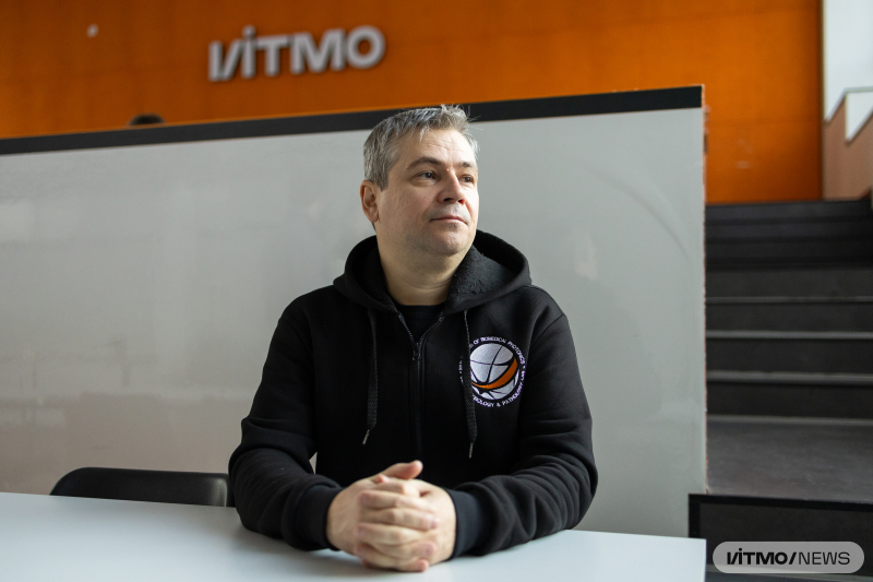
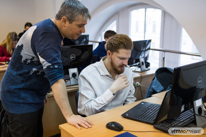
Andrey Dunaev conducts a course for ITMO University students. Photo by Dmitry Grigoryev / ITMO.NEWS
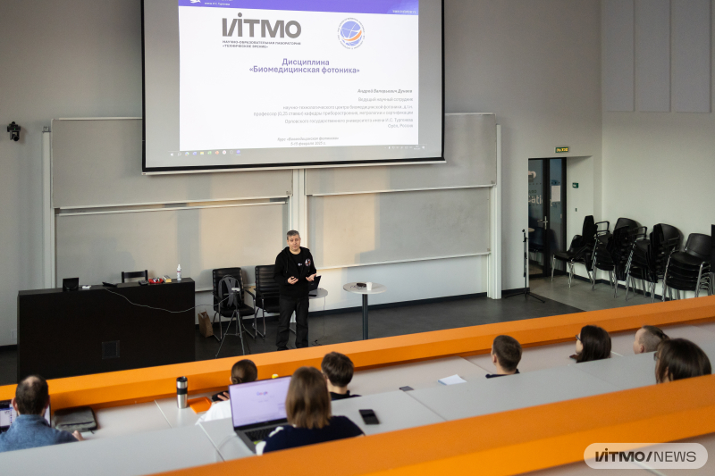
Andrey Dunaev conducts a course for ITMO University students. Photo by Dmitry Grigoryev / ITMO.NEWS
You recently joined the ITMO Fellowship program. How did you learn about it and what made you want to take part?
I’ve known about ITMO for quite some time, since the early 2000s; since 2014, our students have been presenting reports at the Congress of Young Scientists, which is organized by the university. That’s where we learned about videocapillaroscopy – a method of visualizing the movement of erythrocytes through capillaries in tissue at the nail bed. This method can be used to detect various peripheral blood flow disorders, such as in diabetes. Of course, I became interested and got to know those who are working on this at ITMO – professor Igor Gurov and his colleagues, for example, or Maxim Volynsky, the director of the Scientific and Educational Laboratory “Machine Vision,” and others. Since then, we’ve decided to share our experience and ideas. ITMO has more expertise in developing AI technologies and optical systems, while we have more experience in creating medical devices and implementing them in clinical medicine.