To develop new medical materials with specific properties, for example, vascular stents or implants for correcting tissue defects, it is necessary to understand the cellular structure of the substances from which they will be made, as well as the compatibility of these substances with the body’s cells. With this information, specialists can predict whether these materials will achieve the desired effect and be safe for humans.
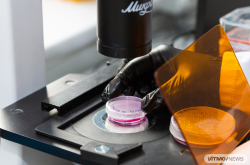
In order to study cellular processes more precisely and track their dynamics, scientists cultivate cells on samples of the material. Next, they segment these cells in microscope images (manually or with specialized software) and identify patterns in their characteristics.
One part of this process looks like this: specialists “trace” the contour of each cell and highlight their nuclei – visually, this is somewhat similar to the paint-by-numbers technique. Thus, a single image is divided into many small fragments with clear boundaries, and as a result, scientists no longer have a shapeless blob with hundreds of cells, but an image where each of them is separated from the others by a clear line. Based on this data, researchers can identify patterns in the shape, size, and other characteristics of the cells and draw conclusions about the compatibility between a given material and cells belonging to a certain tissue type.
Hundreds of such images have to be processed for the conclusions about cell properties to achieve sufficient statistical significance. This typically takes several days. However, with dedicated ML tools, the process can be completed within minutes.
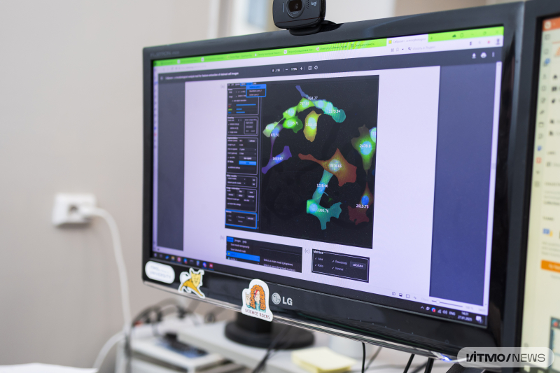
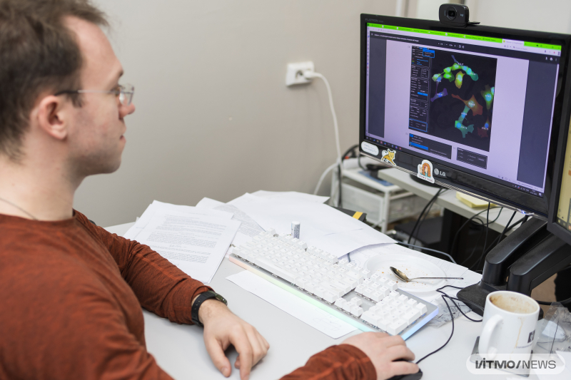
The software for cell structure analysis. Photo by Dmitry Grigoryev / ITMO.NEWS
One of the most popular tools used for this task today is the Cellpose framework, developed by neurobiologists Carsen Stringer and Marius Pachitariu in 2020. Even though now used by biologists and chemists globally, Cellpose can only segment the cells on images – the researchers still collect the statistical data on cell properties manually.
A team of researchers from ITMO have developed a tool that will help automate this step of the process as well. Their software, named Cellpose+, builds on the functionality of the existing framework: it can now automatically collect statistical data on cells’ morphological characteristics from their images in just minutes. Cellpose+ provides information on the total number of cells, their density, area, shape, nucleus location, cell-to-nucleus ratio, symmetry, and, importantly, Voronov entropy that shows how ordered the cells are in the considered plane. Thus, researchers get the data necessary for further analysis of a cell culture and the design of medical materials in minutes instead of days.
“Earlier, to collect statistical data, we had to download images from Cellpose and process them in another program or manually. Now, both of these functions are accomplished by a single tool. First, a neural network trained to segment cells on Cellpose’s considerable library processes the images. Next, our software works with them; the data is collected and classified with feature extraction algorithms. After that, the results are put into separate files in accordance with the indicated cell properties, and these files can be processed by scientists. At each stage, Cellpose+ can be manually controlled to minimize the number of errors,” says Pavel Zun, the head of the research project and an associate professor at the Infochemistry Scientific Center.
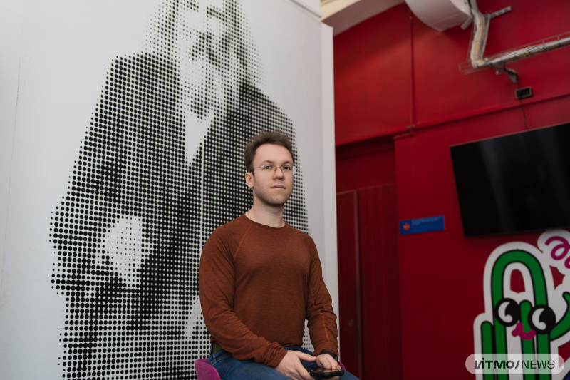
Pavel Zun. Photo by Dmitry Grigoryev / ITMO.NEWS
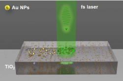
Currently, the developers are testing the program’s capabilities on their own images of polyhydroxyalkanoate polymers. These are biocompatible and biodegradable polymers from which substrates for cells can be made that are structurally and mechanically similar to extracellular structures in the body. Based on the data obtained through Cellpose+, scientists can determine which polymers are most suitable for the survival and further division of a specific type of cell.
In the future, this will help develop a more efficient formula of a polymer that can be used as a biocompatible and biodegradable material for medical implants in regenerative medicine or a chip for delivering drugs to a specific area of the body.
In the future, Cellpose+ can be used for other applications related to the analysis of microscopy images. For instance, it can be useful for tracking the effects of therapy on cancer cells, developing new drugs, detecting anomalous cells in blood tests, and studying the division of a particular kind of cells.
The code written by the ITMO team can be accessed on GitHub, available to any researchers who encounter similar tasks in their work.
The project is implemented within an R&D in AI grant No. 640103 The development of ML-based automated processing and analysis methods of optical and atomic force microscopy images.