Contents
- What is MRI and how does it work?
- Not just medicine: other MRI applications
- MRI development
- MRI in Russia
- How ITMO trains MRI specialists
- What is MRI School
What is MRI and how does it work?
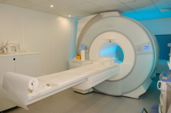
Magnetic resonance imaging (MRI) is an imaging technique that makes it possible to see the internal structure of the body without using ionizing radiation. It is based on the phenomenon of nuclear magnetic resonance: the patient is placed in a strong magnetic field, where the water protons in the body’s tissues begin to absorb radio-wave energy and emit a response at the same frequency. Additional gradient fields ensure that each point in the body produces a response at a distinct frequency, allowing the computer to determine where the signal came from. The response is then detected by specialized receivers and converted into an image.
As the human body is almost 80% water, the signal is produced by nearly all body parts, but in both healthy and pathological tissues the signals have specific attenuation properties; that’s why it’s possible to distinguish between a muscle, a liquid, and a tumor on an MRI scan.
MRI scanner. Credit: jianglianqi / photogenica.ru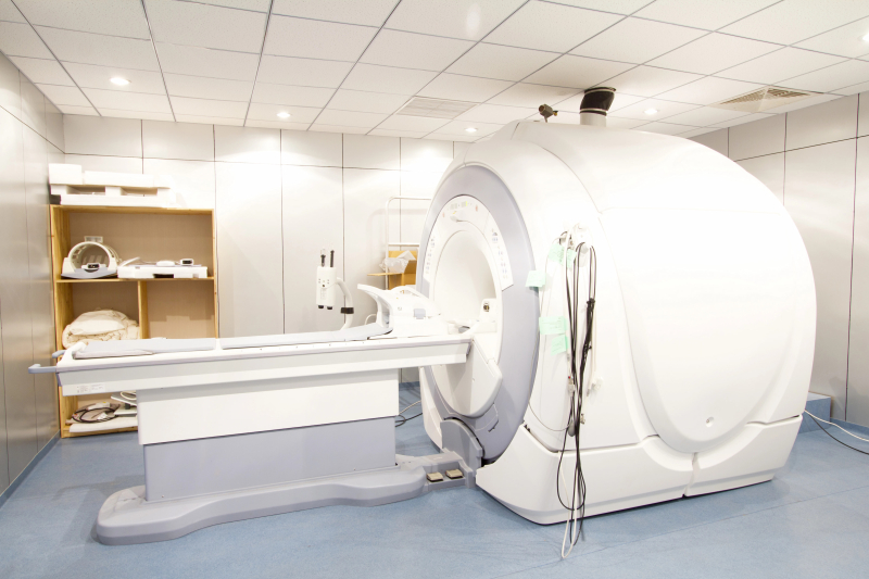
Scanners include three subsystems:
- magnetic creates a constant field; the stronger the field, the clearer the image;;
- gradient (powerful amplifiers increase pulses, while coils change the field along three axes relative to the body, which is why each point has a unique response frequency or initial phase; this allows the computer to tell where the signal came from;
- radio frequency radio-frequency devices generate, amplify, and radiate radio pulses, water protons respond to them, and the receivers catch this response signal.
All three systems are managed by a spectrometer that replays pulse sequences, i.e. preset patterns that activate the systems, at precisely the right moment to adhere to the method and produce a clear image.
Unlike computer tomography and X-ray, MRI doesn’t use ionizing radiation that can damage DNA after frequent exposure. That’s why the method is preferred for many clinical tasks: MRI can be done multiple times. Moreover, it’s one of the most informative visualization methods: it clearly shows soft tissues, including the brain and spinal cord, internal organs, the heart, and the fiber apparatus and cartilage in joints.
However, the procedure has its limitations: it is contraindicated for patients with magnetic metal implants or fragments, as well as implanted devices, because they may shift or heat up in the strong magnetic field.
MRI brain scans. Credit: srikijt / photogenica.ru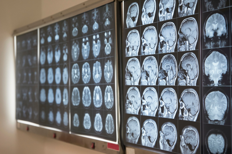
Not just medicine: other MRI applications
Even though MRI is commonly considered a primarily medical diagnostics method, its applications have long traveled outside hospitals. For example, MRI is used to study the internal structure of vegetables and quality of food products. There are also micro MRI machines for eggs that can determine if they are fertilized. Additionally, MRI has found applications in agriculture and oil extraction: it is used to study roots of crops and investigate the structure of rock formations.
In medicine, MRI applications are also expanding. Apart from anatomy, the method can be used to study functional parameters: the rate of water diffusion in tissues, blood flow properties, iron overload, the distribution of infiltrated fatty tissue within healthy tissue, and more. The quality of the resulting image depends on the machine’s settings: by changing them, specialists can focus on different qualities of tissues. This results in new visualization methods such as 4D angiography that makes it possible to study blood vessels in real time (including without a contrast substance) or 3D localized spectroscopy that provides information on the distribution of metabolites in tissues.
All this is to say that MRI is a platform that can be tuned to perform various tasks.
MRI development
Globally, MRI technology is developing in several directions. Among the leading ones is increasing the strength of the magnetic field that patients are placed inside. Hospitals typically use machines with 1.5-3 T fields; however, more powerful systems with fields of up to 7 T are implemented more and more, as they offer higher quality images. Such machines can visualize even separate metabolites, which are substances produced in tissues during metabolism and which can signal the presence of a disease.
Another field of development is accelerating the scanning process: thanks to new electronics, powerful computers, and neural networks the time of the procedure is brought down, while the image quality increases. Another important direction is the creation of portable low-field machines that can be delivered to limited mobility patients.
MRI in Russia
Currently, the Russian MRI market contains almost exclusively imported devices. This creates two problems: first, hospitals depend on imports and international service providers; second, researchers have limited access to equipment needed for the development of new solutions. Clinical MRI scanners have high patient loads, while manufacturers rarely allow scientists to change scanning protocols or introduce custom algorithms and devices.
However, in recent years the situation has seen an improvement. Rosatom has started developing a Russian MRI scanner with a 1.5 T field. The machine will contain a superconducting magnet: at very low temperatures it conducts electricity without resistance, allowing the magnetic field to remain stable without requiring a constant energy supply.
The project team includes ITMO scientists who are developing algorithms for pulse sequence generation, radiofrequency coils and devices, as well as phantoms – test samples that imitate human tissues. The device is scheduled to be completed in 2026. A domestic device will allow hospitals to depend less on imports. Apart from that, introducing the machine will benefit researchers, as well: they will be able to change and create new pulse sequences, introduce and test advanced reconstruction and postprocessing methods, as well as suggest new engineering solutions. This means that the demand for MRI specialists is about to increase.
How ITMO trains MRI specialists
Specialists who have a grasp of MRI physics and are capable of working with equipment and developing new radiofrequency devices, as well as image acquisition and processing methods, are trained within ITMO Bachelor’s program Wireless Technologies, which offers a special track called MRI Devices and Methods. This program by the Faculty of Physics is the only one in the country that focuses specifically on MRI technology development and not medical equipment in general. During their studies, students engage in the university’s projects, gaining practical experience along with theoretical knowledge.
Graduates can work on Rosatom projects, at service companies that install and maintain MRI scanners, as well as at companies that develop medical software and equipment.
Moreover, ITMO has several laboratories whose members have spent over a decade working with MRI technologies. The researchers develop flexible substrates for imaging a fetus’s brain in the womb, universal wireless coils for scanning with MRI systems from different manufacturers, new types of antennas for ultra-high-field MRI, and portable scanners that can be brought directly to a patient’s hospital room. Additionally, the scientists are working on new image processing algorithms: they help accelerate image acquisition and can automatically measure the heart’s parameters – for instance, assess scar tissue areas after a heart attack.
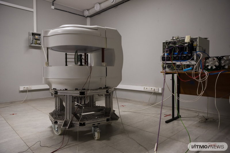
Prototype portable MRI scanner. Credit: Dmitry Grigoryev / ITMO NEWS
What is MRI School
Globally, MRI research is coordinated by the International Society for Magnetic Resonance in Medicine (ISMRM), which introduces international safety standards and organizes major annual conferences. The organization has members in over 65 countries, including Russia. However, at the moment Russia doesn’t have its own internal community of MRI physicists and engineers, which is why specialists from different cities and organizations rarely have the chance to communicate. In order to help establish such communication, ITMO has been annually organizing the MRI School and Conference for four years. This year, the event is supported by the Vladimir Potanin Foundation. Speakers from Rosatom’s research department have also joined the event. Participants attend lectures by leading specialists in the field and work on projects in teams.
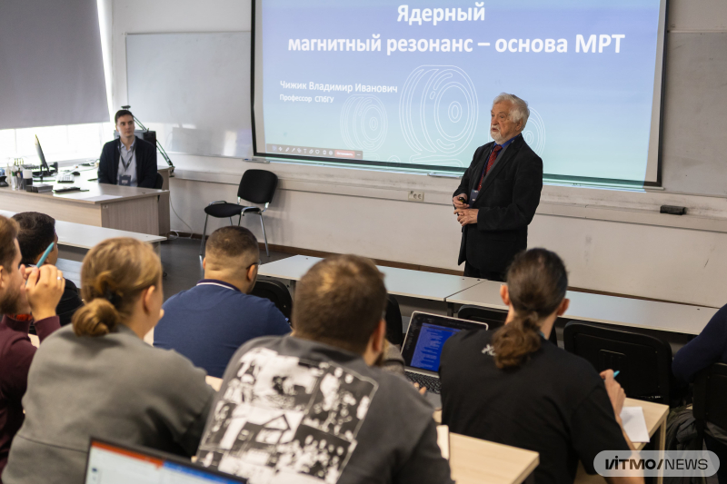
MRI School. Credit: Dmitry Grigoryev / ITMO NEWS
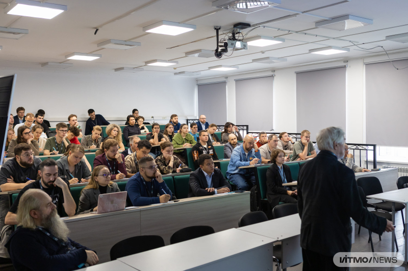
MRI School. Credit: Dmitry Grigoryev / ITMO NEWS
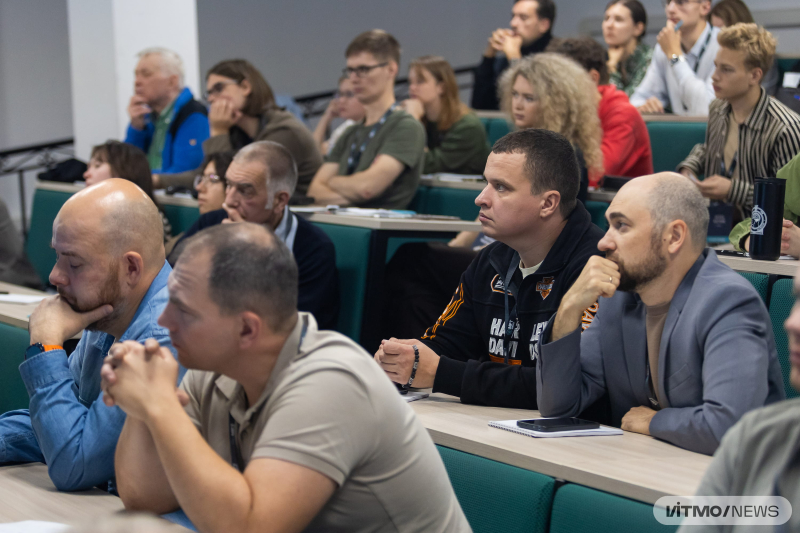
MRI School. Credit: Dmitry Grigoryev / ITMO NEWS
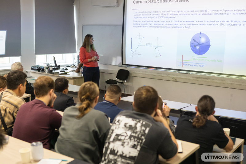
MRI School. Credit: Dmitry Grigoryev / ITMO NEWS
At the event, participants could discuss their studies, solutions, and projects with experts from academia and industry, including MRI device manufacturers and service companies.
Within the school, students are introduced to the basics of the technology and can choose the direction of their future studies: either equipment or methodology. Experienced engineers, on the other hand, can explore the relevant approaches in practice. At the end of the school, students take a final test; those who score over 70% receive a continuous professional development certificate.
This year, the school brought together over 70 participants from all across the country, from St. Petersburg to Vladivostok. Around half of the participants are students of medical physics, biomedical engineering, and biophysics. The other half are engineers working at hospitals and service companies, as well as organizations developing medical equipment.