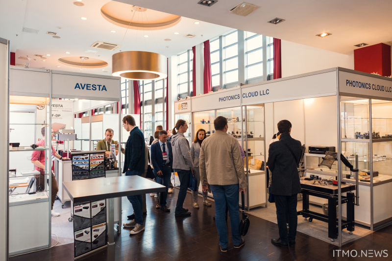
This summer, the International Conference Laser Optics (ICLO 2022) was held on June 20-24 in a hybrid format and featured symposiums, plenary, parallel, and poster sessions. The participants discussed topics in the fields of laser physics, optics, and photonics: semiconductors, powerful and free electron lasers, quantum optics, non-linear photonics, biomedical applications, etc. Moreover, participating researchers had the chance to join the 7th International Prokhorov Symposium in Biophotonics and a round-table discussion in honor of the 100th anniversary of birth of Nobel laureate Nikolay Basov, as well as attend international summer schools and present their works at the Lasers and Photonics exhibition.
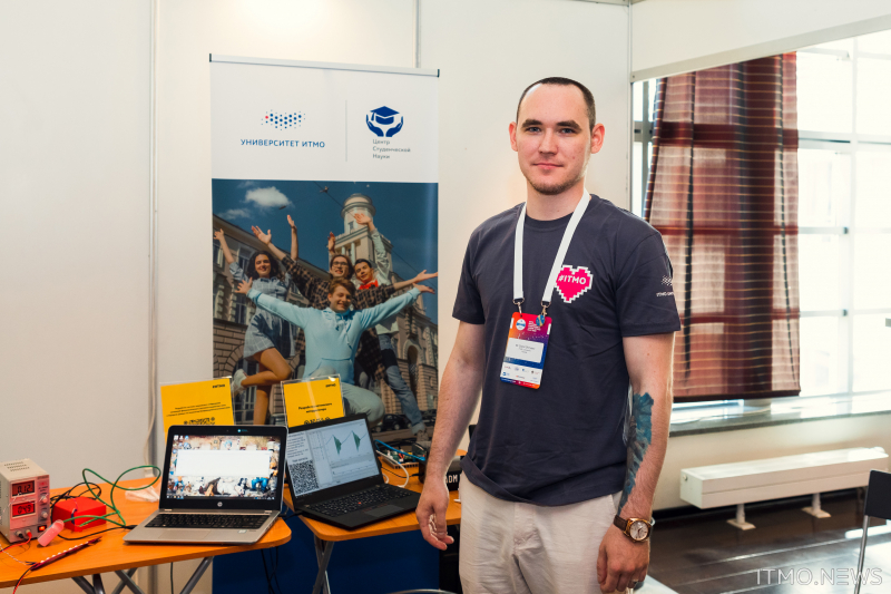
Four young researchers from ITMO – PhD students and research associates – took part in the exhibition. They showcased a portable two-channel microscope, an optical interrogator, a semiconductor photodiode sensitive in the UV range, and an online patient monitoring system.
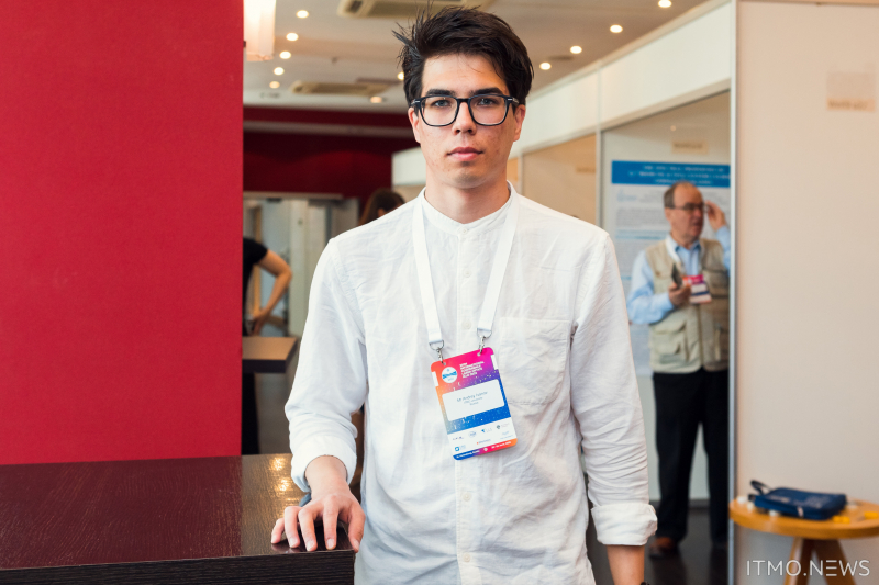
“By exhibiting their work at such events, students get another opportunity to receive feedback that will come in handy when they defend their theses. Our participants are young researchers, including heads of various research and engineering laboratories, who as students managed to assemble teams to implement their ideas. All of them spearheaded their own projects and won the contest for practice-oriented projects held annually at ITMO, which allowed them to receive up to 2.5 million rubles in funding. The winning teams have to develop a full-scale prototype along with technical documentation for further scaling and commercialization of their projects,” explains Natalia Karmanova, a first-year PhD student at the Faculty of Secure Information Technologies and a representative of ITMO’s Center for Student Science, Conferences, and Exhibitions.
Natalia Karmanova. Photo by Dmitry Grigoryev, ITMO.NEWS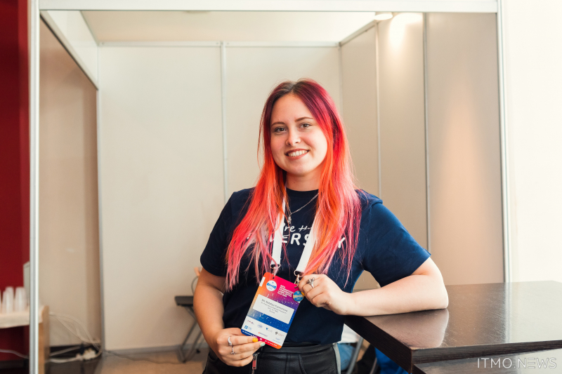
Zooming in on a wider picture
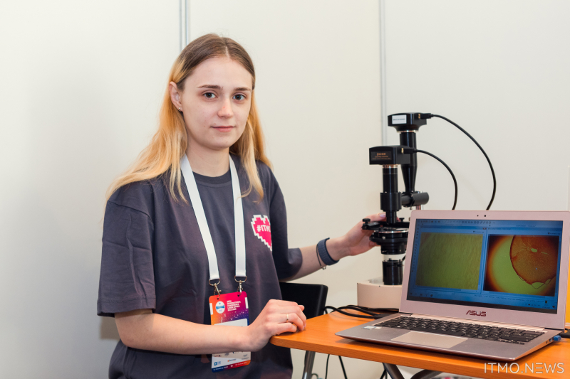
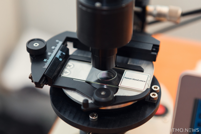
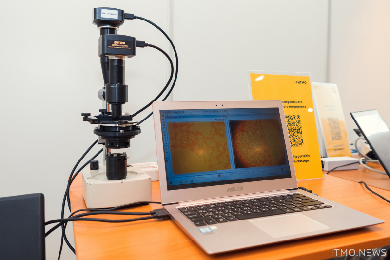
In late March 2020, the team headed by Anastasia Kozhina, a third-year PhD student at the Higher School of Engineering and Technology, won the grant for their practice-oriented project that aimed to develop a portable two-channel microscope. A special feature of the device is that while one of its lenses allows a full overview of the biological sample, the second lens can present a single area with a 40x magnification. Moreover, the microscope makes it possible to analyze samples right after their collection without the need to transport them to a lab.
After two years of work, the researchers progressed through the theoretical part of their work and were able to present a real-life prototype at the exhibition. Now, the device allows viewing a sample in two modes at once and tracking changes in real-time without additional refocusing – users simply need to shift the microscope’s stage. The device’s counterparts available on the market require researchers to constantly change lenses and refocus them in order to identify and analyze areas of interest. Apart from taking more time, by requiring constant refocusing on a changing sample, such as blood, these steps may lead to losing the area of interest that changed during such “shifting of gears.”
“We are planning to polish the engineering and software aspects of the microscope. First of all, we want to improve some of its optical features to ensure images of higher resolution. Second, currently the device doesn’t allow us to see which part of the sample we are zooming in on, which means that for now we are left to guess its exact location in the sample. In the future, we are hoping to develop an app that would allow users to not only analyze the sample but also pinpoint the zoomed-in area seen in one lens on the broader picture seen in the other,” shares Anastasia Kozhina.
Currently, the team is testing and improving the microscope’s features so that in the future the device can be offered to hospitals, clinics, and veterinary clinics for testing biological samples and in particular identifying parasites in blood.
Tracking oscillations
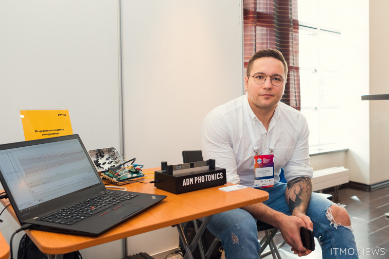
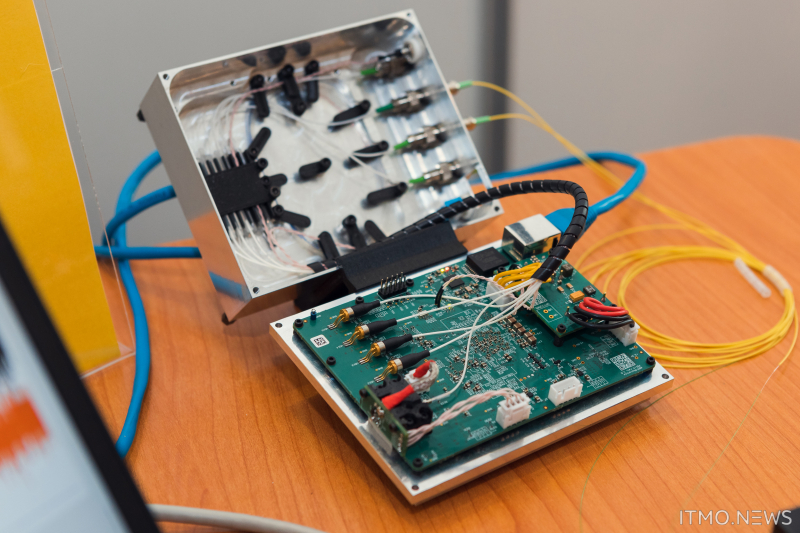
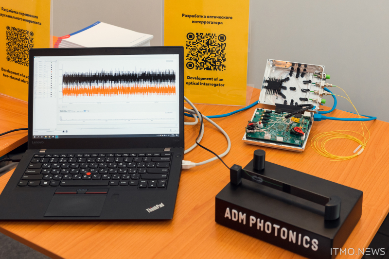
The team headed by Sergey Volkovsky, an assistant at the Higher School of Engineering and a researcher at the Research Center of Light-Guided Photonics, developed an optical interrogator. This is a device that measures the physical parameters of various objects, for instance, their deformation, temperature, pressure, and the like.
“We received an order from representatives of the aviation industry, who needed a device capable of tracking the deformation of helicopter blades in real time. These blades are quite expensive and are currently replaced after they have performed for a specified amount of time, meaning that they aren’t inspected for defects. However, not all blades wear out over the same amount of time and that’s why it became necessary to track their deformation level and only replace them when it is needed. This would make it possible to not only cut down on the associated costs but also increase flight safety,” explains Sergey Volkovsky.
Here is how the device works. The optical interrogator contains a laser with a controlled wavelength and is connected to special sensors, fiber Bragg gratings. These sensors are subjected to the laser’s radiation, part of which they reflect back to the interrogator, which extracts information about the sensors. This information is displayed for the user in real time via the digital interface. At the core of this is a property of Bragg gratings to change their oscillation cycle when stretched or compressed, which in its turn leads to changes in their reflection spectrum registered by the interrogator.
One of the first prototypes of the device has already been tested on the rotor of a helicopter. Its blades had built-in arrays of Bragg gratings, which allowed the interrogator to perform its calculations, eventually wirelessly transmitting the information about identified defects in the blades to the operator. According to the researchers, the device is currently at a pre-production stage and can become a production sample after a number of small fixes.
The new interrogator has a wider field of applications, including construction, where it can be used to monitor the integrity of objects of infrastructure, plumbing, railroads, and buildings. Moreover, it can even be used in space to monitor temperature shifts in those conditions when other sensors cannot be used.
UV protected
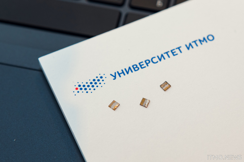
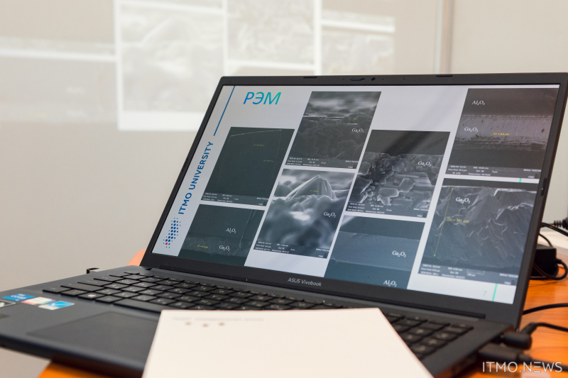
Various photodiodes made of semiconducting materials can be found on the market, including wide band ones, based on silicon carbide, nitrides of third group metals, and other similar materials. However, their sensitivity in the deep UV range is much lower, which is why they cannot verifiably detect radiation in this range. This can be fixed. For this purpose, the team of Andrey Ivanov, a first-year PhD student and an engineer at the Institute of Advanced Data Transfer Systems, developed a light-sensitive semiconductor device, namely, a photodiode with a Schottky barrier, based on gallic oxide (Ga2O3).
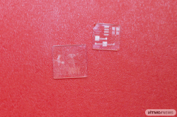
At the exhibition, the team showcased one of the diode’s components, a device structure of gallic oxide with an indium ohmic conductor and a gold Schottky contact. The structure will later be located inside the completed device. When subjected to deep UV radiation, photons in the active field transmit their energy to electrons in the valence band and make them go through the band gap. This results in a more intense photo-induced current than in the dark, thus signifying the presence of UV radiation. If, however, the active field is subjected to visible light or infrared radiation, the resulting energy will not be enough to produce visible changes in the device’s volt-ampere properties.
Currently, the researchers are testing photodiodes of their own design as part of the third stage of their project aiming to improve their photosensitivity. Moreover, the team is developing the body for the corpus structure.
“Some factors, such as deep ultraviolet radiation, can be harmful to people, that’s why we’ve developed a device that will track this radiation. It can be used in a number of fields. For instance, in healthcare and biology for helping people with various skin conditions, in detecting high-voltage charges in power lines or flames at hazardous production facilities, in UV spectroscopy, communication systems, ozone layer monitoring, and even astronomical observations,” says Andrey Ivanov.
A wristband to save lives
Daniil Shiryaev, a second-year PhD student at the Institute of Advanced Data Transfer Systems and a junior researcher at the Laboratory of Single-Photon Detectors and Generators, together with his colleagues developed a system for automated monitoring of patients who are in a state of coma or early recovery stages. Currently, there exist several non-invasive and invasive methods to track changes in the states of such patients, however, the device developed by the team stands out by being able to visualize the acquired data.
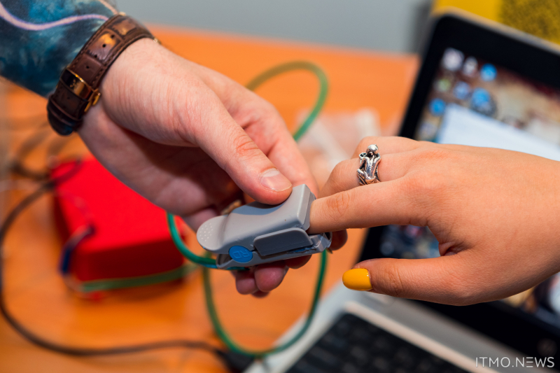
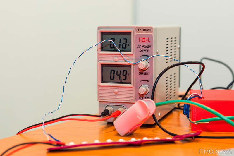
According to the suggested design, medical sensors (a thermometer and a pulse oximeter) are put on a patient’s finger to measure three parameters, namely, heart rate, body temperature, and blood oxygenation level. Raw data is sent to the control panel, where it is processed. There, the algorithm detects whether the acquired value falls within the norm. In case it does, the control panel sends corresponding signals to the server and the wristband, which keeps the LED band in it green. Otherwise, the LED band will change its color depending on the parameter that’s out of order and start blinking in a wave-like motion. At the same time, the attending physician will receive a warning message on their phone.
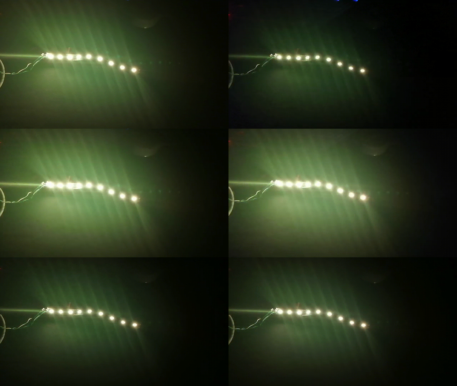
Changes in color and pulses of LEDs on the wristband in reaction to the patient's temperature changes. Video courtesy of the project's authors
At this stage, the researchers already have a mock-up of the device and are in the process of compiling the engineering documentation for a test sample. Assembly and testing of the sample at the Polenov Neurosurgical Institute are scheduled for this autumn.
“Bedside monitors are typically used at hospitals to monitor patients. Such monitors are mainly located in intensive care units, however when the patients get allocated to the general ward it is still important to continuously monitor their state – but there are no monitors there. Our wristband will make it possible to tell which measure is out of order and this way save time on making a decision for each patient. We are also planning to develop an app that will enable patients to keep monitoring their health after their discharge from the hospital, while also giving access to this data to their close ones,” shares Daniil Shiryaev.
Apart from that, the research team will be connecting an encephalography cap to the wristband in order to enable the device to analyze the brain’s bioelectric activity and develop software that will accelerate preprocessing of such data, helping to visualize the results of the analysis. The cap will not only increase the observations’ reliability but will also be useful for studying the underlying processes of chronic disorders of consciousness.
