In collaboration with Active Photonics, ITMO researchers have developed a device that, unmonitored, can produce highly accurate results in microbiological and cell experiments. It combines two methods for studying substances: atomic-force microscopy and Raman spectroscopy. Experimental data are automatically analyzed with built-in software.
Using a nanometer-scale tip, atomic force microscopy “feels” the surface of a material and constructs a 3D map of its topography. Raman spectroscopy, in turn, determines the chemical composition of a substance by analyzing how it scatters laser radiation. Typically, these methods are implemented using two separate tools, which is expensive and time-consuming. However, combining both methods in a single setup enables simultaneous observation of both the composition and shape of a material, increases the accuracy and quality of the resulting data, and ensures that the investigation is conducted in the desired local area.
The new device is set up in the portable chemical “cube” for cell and bacteria experiments – a fully automated mini lab built at ITMO in 2023. Inside, there is a robotic hand that acts like a lab assistant and automates a part of the experiments – thus, increasing their accuracy, including via the possibility of multiple automated repetitions.
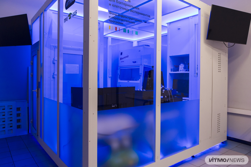
The mini laboratory at ITMO. Photo by Dmitry Grigoryev / ITMO NEWS
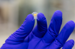
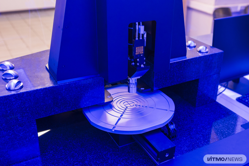
The system operates as follows. A robotic manipulator places a plate reader containing samples of the substance (in this case, cells or materials) onto a movable platform. On this platform, the plate is moved into the scanning area: the atomic force microscope collects information about the surface topography, while the spectroscope gathers data about the material’s properties and composition. The built-in software analyzes the obtained information, which is then made available to researchers.
ITMO scientists collaborated with the company Active Photonics on the setup – their partners asked for a system that would combine the two matter scanning methods. This will facilitate the development of new materials, increase replicability, and decrease the probability of errors in data analysis.
“With this device, it is possible to measure the mechanical properties of the cell membrane (such as stiffness, elasticity, and adhesion), perform chemical analysis, monitor the quality of cell cultures, and study interactions between cells and biomaterials. All of this helps investigate the properties of materials and the specifics of their modeling, providing insight into how these materials interact with biological systems. This information is valuable for the development of materials for biological applications – for example, in creating biocompatible coatings for implants, drug delivery systems, and in tissue and organ cultivation,” shared Alexey Meshkov, shared the main developer of the project and a researcher at ITMO’s Infochemistry Scientific Center.
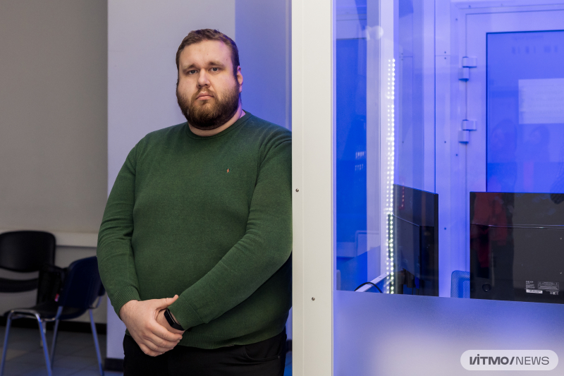
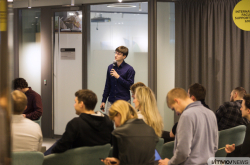
Alexey Meshkov. Photo by Dmitry Grigoryev / ITMO NEWS
ITMO scientists and students working on the automation of chemical technologies – including researchers from ITMO’s Laboratory of Atomic Force Microscopy – will operate the new device. In the portable laboratory, they are already searching for and studying new cryoprotectants to discover new molecules, developing robotic platforms to monitor the growth and metabolic activity of microorganisms in real time, and investigating the influence of nanostructures in polymeric, hybrid, and biodegradable materials on cell growth. The latter studies will help find new solutions for regenerative medicine. For example, researchers are learning to program the behavior of cardiomyocytes (heart muscle cells) using biodegradable polymer products – with the potential to scale these developments toward creating a fully functional artificial heart.