Polyhydroxyalkanoates, or PHAs, are biodegradable polymers derived from bacteria. PHAs are biologically compatible with living cells and tissues and degrade into non-toxic byproducts inside the body. All this makes them the ideal candidates for biomedical applications – for instance, implants, sutures, and scaffolds for tissue engineering. However, simply obtaining a biocompatible material and a medical product is not enough; cells need to successfully attach to and grow on its surface. This is subject to the roughness of the surface at micro- and nanoscales. Consequently, the surface profile of the material is crucial for biomedical applications.
Researchers from ITMO University and the Federal Research Center “Krasnoyarsk Science Center of the Siberian Branch of the Russian Academy of Sciences", alongside their colleagues from the Netherlands and the USA, have determined the optimal thickness and surface topography of PHAs that enhance attachment and growth of cells. The films were produced using a special biodegradable polymer of the PHA family chosen due to its great processability, biocompatibility, and cost-effectiveness for large-scale biosynthesis. Four solutions of various concertations were used during the fabrication – the higher the concentration of the solution, the thicker and rougher the film was. For the next stage, the researchers cultured myoblast C2C12 directly on the polymer surfaces. These cells are muscle tissue precursors and are among the most convenient and common objects for biomaterial testing.
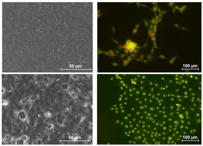
Images of films made of PHAs (left) and myoblast cells that grow on the obtained films (right). Credit: Federal Research Center "Krasnoyarsk Science Center of the Siberian Branch of the Russian Academy of Sciences"
During their study, the researchers had to figure out how to effectively measure if a surface is suitable for cell growth. Typically, scientists do this manually: they count cells, take measurements, and describe the topography. But this method is time-demanding, subjective, and poorly reproducible. For that purpose, the team developed a reproducible algorithm that automates the process: namely, it segments cell images and generates metrics for both surface profile and cells. The algorithm also connects all stages into a single chain – from film fabrication and surface characteristics to data on cell behavior. As noted by the developers, the technology eliminates random guessing by enabling a targeted selection of parameters and is applicable for all types of human cells when developing new polymer-based implants.
“For this study, our research team from ITMO’s Infochemistry Scientific Center developed an approach that automatically evaluates how cells interact with polymer film surfaces. First, a convolutional neural network (CNN) segments cell images captured by a fluorescence microscope, then the quality of this segmentation is verified. The data obtained is used to determine the core characteristics of cells: their shape, size, and other morphometric features that we compare with the surface properties we obtained from the images taken using atomic force microscopy (AFM) and scanning electron microscopy (SEM). Thus, we identify the parameters of surfaces that strongly correlate with the biocompatibility of samples,” notes Pavel Zun, an associate professor at ITMO’s Infochemistry Scientific Center and the leader of the research team in biomedical computing at ITMO University.

Pavel Zun. Credit: Dmitry Grigoryev / ITMO NEWS
Thanks to the algorithm, the researchers were able to record how cells behaved on films: how they attached and grew and how efficiently the film supported these processes. The study showed that cells attach better to thick films with rough surfaces. A crucial part is played by various features on the surface that cells can adhere to. Thin films made from weak solutions have a mostly smooth surface and therefore are harder for cells to attach to. Whereas thicker films are filled with irregularities and depressions that promote greater cell attachment, as well as higher growth and activity of cells.
“The solvent evaporates more slowly in films made of more concentrated and viscous polymer solutions. This has to do with the fact that higher viscosity dampens the motion of solvent molecules from the depth to the surface. This means that the polymer crystallizes more slowly, as well. The delayed crystallization is accompanied by the formation of larger and more rigid structures, which create a more developed and rougher film surface. Cells are ‘seeded’ onto the film, just as plants in a garden bed. The roughness of this ‘bed’ determines how well the cell will ‘sit’ and multiply on it. The study will prove useful for the engineering of biomedical surfaces based on PHAs and other polymers. The results can be used in tissue engineering to produce scaffolds that promote cell growth and formation of new tissues – for example, for the reconstruction of muscles, bones, cartilage, and blood vessels – as well as in regenerative medicine for implants that need to faster integrate with patients’ own cells, form new tissues at the injury site, and avoid rejection,” comments Tatiana Volkova, DSc in biology, a professor, and the head of the Institute of Biophysics (Siberian Branch of RAS) and the Department of Biotechnology of the Siberian Federal University.
Written jointly with the Federal Research Center "Krasnoyarsk Science Center of the Siberian Branch of the Russian Academy of Sciences"