Magnetic resonance imaging (or MRI) is considered one of the most advanced methods for diagnostics available to modern medicine. MRI scanners can detect problems in internal organs and tissues, joints, or the blood system, as well as identify tumors and much more. Clinicians in various fields send their patients to undergo an MRI scan before finalizing their diagnosis.
For breast cancer – the most common cancer type in women – the results of an MRI scan may not only be crucial in confirming the diagnosis but also save lives, as early detection of the tumor can almost guarantee the full recovery of a patient.
“Compared to an X-ray or ultrasound examination, an MRI scan allows us to precisely localize the tumor, which affects the accuracy of the diagnosis,” explains Alena Shchelokova, first co-author of the paper and research associate at ITMO’s Faculty of Physics and Engineering. “MRI is especially relevant for young women with dense breast tissue or breast implants when mammography is often incapable of distinguishing between healthy and cancerous tissues.”

However, despite the possibilities open to medical specialists by MRI, this method is rarely used to produce breast scans because apart from the scanner itself, this procedure requires additional specialized equipment. As this equipment is rather expensive, not every medical center can afford it. As a result, the standard set of MR scanners is used for diagnostics, and the images obtained after scanning are insufficiently informative for making a diagnosis, and small details may be missed entirely. To compensate for this, clinicians often resort to an increase in scan time due to its multiple repetitions, which, however, leads to the fact that the throughput even in high-tech MRI rooms becomes very low, and patients experience discomfort from a long time inside the scanner. As a result, only women in the increased risk group receive the necessary examination procedure, and for young patients without hereditary predispositions, this procedure remains practically inaccessible.
A question of equipment
The operation of an MR scanner is based on the excitation of protons, the nuclei of hydrogen atoms, using radio waves and recording their response signals. For this, a strong constant magnetic field is created inside the scanner in which the protons pass into a higher energy state. Being in this state, they can receive and give energy in the form of radio waves familiar to everyone. Collecting radio signals from protons inside the diagnosed area, makes it possible to construct a detailed anatomical image of a selected part of the human body. Moreover, in different organs and tissues of the body, this radio emission will differ due to the inhomogeneous density of protons and some other characteristics of biological tissues, which makes it possible to obtain a contrast between them in the image, and therefore to quickly identify signs of diseases.
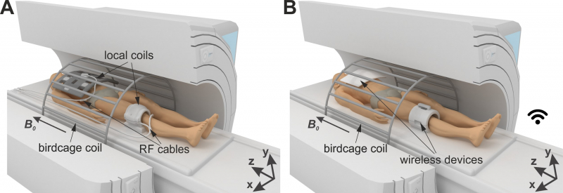
The heart of a typical clinical magnetic resonance imager is a large transceiver coil (in MRI, specialized antennas are called “coils”). This birdcage-type body coil excites protons in the human body. In terms of its size, it is comparable to the tunnel in which the patient lies during the examination. It allows the creation of radiofrequency fields of high spatial uniformity so that most of the protons receive their equal portion of energy, and the output image is free of any dark spots. At the same time, it is not suitable for receiving weak responses from nuclei. For this, other, smaller coils of various configurations are used, optimized for a particular region of the body (head, knee, back, etc.).
“There are two key processes in MRI – excitation and signal reception,” expounds Alena Shchelokova. “The birdcage coil cannot provide an MR image with sufficient contrast and signal-to-noise ratio because it is large and located far from the specific area of interest. That’s why small dedicated coils are used as receiving antennas for each organ or body part. Usually, medical centers are only equipped with a limited number of these local coils, and the breast coil is rarely found among them. As a result, a breast scan would simply be of such low quality that diagnostics would be impossible.”
Ceramic resonator
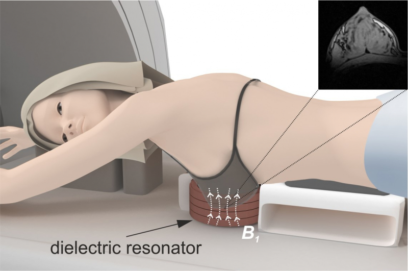
A group of Russian researchers headed by ITMO University scientists has proposed a different approach to the problem. The novel concept of targeted clinical MRI applied to breast imaging is based on a local redistribution and passive focusing of the radiofrequency magnetic field of the large body coil through electromagnetic coupling with an additional dielectric resonator surrounding the target area. This approach makes it possible to avoid using a dedicated receiving coil for the breast.
“Let’s introduce a photography analogy – just imagine that instead of taking a panoramic picture, you are zooming in and focusing only on the one detail that interests you,” continues Alena Shchelokova.
The device itself is a small cylindrical resonator made from a unique type of ceramics developed by the St. Petersburg company Ceramics Co., Ltd.
“Modern ceramic materials are multicomponent complex polycrystalline substances. We create new ceramic materials based on our experience and the analysis of preliminary research conducted in the field that uses modern physical-chemical analysis methods and metrology. We can control the materials’ properties and develop ceramics for specific purposes, especially in electronics. Moreover, we not only create the materials themselves but also use them to develop products,” says Elizaveta Nenasheva, CEO of Ceramics Co.

The ceramic material in the composition of the resonator has a high value of the dielectric permittivity, which makes it possible to reduce the speed of propagation of electromagnetic energy inside the material, and, consequently, to focus it by giving this material a specific spatial geometry. More importantly, this material has low dielectric losses, allowing specialists to avoid a significant effect on the amplitude of electromagnetic waves without leading to their rapid decay.
Being tuned to the frequency of the scanner, the resonator localizes and amplifies the magnetic component of the radiofrequency field in the breast. At the same time, the excitation and reception of the signal are still carried out by a large body coil, which, by the way, is a part of any clinical scanner.
“Our device electromagnetically couples to the birdcage coil and allows us to improve its capabilities by focusing the magnetic field in the area of interest,” explains Alena Shchelokova. “It doesn’t have to be connected via radiofrequency cables to the scanner; it can simply be placed on the scanner table with the patient lying on it in order to get high-quality MR images of the breast.”
Comparison to analogs
Thanks to the fact that the entire radiofrequency field, which was supposed to affect the body during the scanning procedure, is focused in the area of interest (in the breast) by the resonator, it becomes possible to significantly reduce the power of the radio signal required to excite protons. Accordingly, MRI examination becomes even safer for patients in terms of potential heating of body tissues.
The device itself is also cheaper than the specialized receive coil for the breast provided by well-known manufacturers. Additionally, it doesn’t need to be connected to the scanner with a complex system of wires – it is completely wireless and doesn’t have so many fragile parts. Moreover, the resonator is compatible with all models of standard clinical scanners (produced by Siemens, Philips, General Electric, etc.). In contrast, the standard receive coils need to be purchased from the same manufacturer that produced the scanner.
The researchers carried out a number of tests on volunteers with various body shapes. As a result, the test using the new ceramic resonator was able to provide images comparable in their quality to those produced by the most advanced specialized native coil – all this with the increased radiofrequency safety of the scanning.
“MRI is the most sensitive method in diagnosing tumors in the breast. However, the specificity of the standard scanning methods is limited, which leads to a large number of false-positive results. Furthermore, the scanning itself takes a rather long time, which limits the use of MRI in breast cancer screening. Lately, there is a lot of research aiming to find optimal and short methods for breast MRI specifically for their application in screening. And developing a ceramic resonator that focuses the scanner’s visualizing capacity on a specific area of interest is also a step in the direction of breast cancer screening with MRI. The resonators make the breast MRI scan shorter and more accessible, as well as make the MRI visualization more specific by using narrowly specialized scanning methods that are unavailable for standard configuration scanners due to safety regulations,” comments Anna Andreychenko, a research fellow at ITMO’s Faculty of Physics and Engineering.
From research to commercial product

“We’ve been conducting research in the field of applying new artificial materials in MRI scanning for more than eight years. This research started with solving a fundamental task that led us to the highly interesting multifaceted world of MRI. For the last couple of years, we’ve been focusing on transitioning from fundamental research to developing actual products that could be used in Russian medical centers right now. And we’ve succeeded! The resonator for breast scanning is a unique and very useful solution, and I am sure it is to enter the market very soon. We are open and looking for a partnership with all healthcare system representatives, as well as business professionals and investors ready to help us promote this product on the Russian market,” says Irina Melchakova, head of ITMO’s International Laboratory “Applied Radioengineering” and staff member at the Faculty of Physics and Engineering.
This project was conducted with the Vreden Russian Institute of Traumatology and Orthopedics (St. Petersburg), the All-Russian Center of Emergency and Radiation Medicine named after A.M. Nikiforov (St. Petersburg), Ceramica Co., Ltd. (St. Petersburg), the Research and Practical Clinical Center of Diagnostics and Telemedicine Technologies (Moscow).
Reference: Alena Shchelokova, Viacheslav Ivanov, Anna Mikhailovskaya, Egor Kretov, Ivan Sushkov, Svetlana Serebryakova, Elizaveta Nenasheva, Irina Melchakova, Pavel Belov, Alexey Slobozhanyuk & Anna Andreychenko, Ceramic resonators for targeted clinical magnetic resonance imaging of the breast, Nature Communications, volume 11, article number: 3840 (2020).