MRI scanning is used to diagnose brain pathologies – such as strokes, tumors, or neurodegenerative diseases. However, conventional machines require additional infrastructure. First, 1.5T or 3T MRI scanners need a custom-built, shielded room, also known as a Faraday cage, to protect sensitive radioelectronic equipment from external interference. Furthermore, the built-in electric superconducting magnet needs to be continuously cooled with liquid helium, which gradually evaporates to the atmosphere. To avoid the loss of expensive gas, cooling systems or chillers are usually placed outdoors; the chillers and the magnet are connected by cryogenic pipelines, through which helium circulates at ultra-low temperatures (below -150°C).
Because stationary scanners cannot be moved to other rooms, bedridden patients at intensive care units cannot undergo MRI scanning as it would put them at heightened risk for various health complications. Moreover, a single MRI machine can cost more than 100 million rubles, plus 10-15 million rubles yearly for its maintenance. Therefore, MRI scanners are more common in large cities, while residents of smaller cities are often left without the necessary diagnostics.
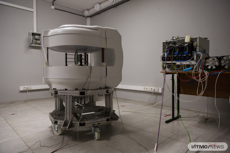
The experimental prototype of the portable MRI scanner. Photo by Dmitry Grigoryev / ITMO NEWS
Initially, MRI scanners employed a low field, but then the field strength started to increase: the stronger the field, the higher the signal-to-noise ratio and the sharper the image. However, as the power grew, so did the size of the equipment, its cost, and insulation requirements. Now, with the advent of low-noise electronic devices and highly-efficient processing algorithms, it has become possible to return to a low magnetic field and create a compact machine on a permanent magnet.
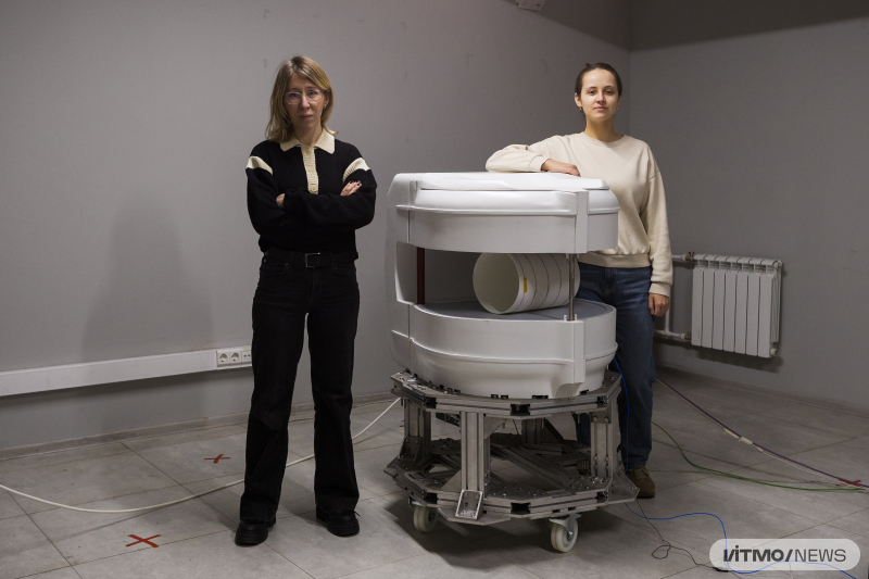
“Unlike traditional scanners, portable ones can be moved to the patient, rather than having them travel to the scanner themselves. To make it autonomous, we looked past a superconducting magnet that needs liquid helium and a complex cooling system; instead, we opted for a permanent, low-field magnet. Since it doesn't need any cryogenic infrastructure, the setup is compact and can operate in any room that has a regular 220V power socket. The scanner can be easily rolled up to a bedridden patient; for examination, the head of the patient needs to be placed into the scanning area,” says Anna Hurshkainen, a senior researcher at ITMO’s Faculty of Physics, who led the scanner’s hardware development.
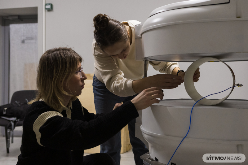
The experimental prototype of the portable MRI scanner. Photo by Dmitry Grigoryev / ITMO NEWS
The portable scanner includes four key modules. The magnetic system generates a permanent field and polarizes hydrogen nuclei in the patient’s tissues. The radiofrequency system excites spins (a collection of particles with their own magnetic momentum) and receives the resulting signal of the nuclear magnetic resonance. The gradient system encodes the signal and creates a 2D image. The spectrometer acts as a computational core: it generates pulses, receives and processes data, and then transmits it to the operator console. As of now, the scientists are about to finalize the integration of all these four subsystems – the next prototype will feature an updated magnetic system.
The ultralow field of the portable MRI (70 mT) enables it to be used to examine patients who cannot do high-field scans. According to the state standard ГОСТ Р 59093-2020, systems up to 0.5T are safe to use in people with metallic implants; the weak field does not heat or displace the metal and causes no harm to the patient. This makes scanning possible for patients with shrapnel or implants, as well as those on mechanical ventilation. The scanner might be set up in a hospital room, an ambulance, or even at a veterinary clinic where it is not possible to construct a Faraday cage. For that, the researchers will need to develop specialized radiofrequency coils that will take into account anatomic features and provide a tight fit for quality signal reception.
The system is equipped with an interference suppression filter based on machine learning and neural networks. Since the scanner uses a low field, the useful signal it receives from tissues is small and can be easily drowned out by any interference. While traditional machines have a custom-built room to address this issue, the ITMO-developed scanner solves the problem differently: additional antennas on the surface capture electromagnetic interference in the environment and the algorithm separates it from the patient-derived signal in the real time. The technology adjusts to both the environment and the patient: body tissues like muscles, fat, and blood exhibit different radio-wave conductivity, causing the patient’s body to act as an antenna. The neural network continuously analyzes and compensates interference to ensure a clear signal. AI does not work with MRI images; it uses initial radio signals to generate the image. The algorithms clean the signal before reconstruction, leaving no room to additive noise in images.
“We can use numerical simulations to generate synthetic MRI data that match the data acquisition method we use in the scanner – which is based on pulse sequences. Then, we add the noise, interference, and distortions that occur in the same machine due to the low-field environment and external noise and thus obtain a pretraining dataset for the neural network. By showing both signals to the network, we train the algorithm to separate useful data from noise,” notes Ekaterina Brui, a senior researcher at the Faculty of Physics, who led the development of methodology and algorithms.
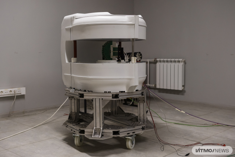
The experimental prototype of the portable MRI scanner. Photo by Dmitry Grigoryev / ITMO NEWS
According to the engineers, a mass-produced portable scanner will cost around 25 million rubles, which is nearly five times cheaper than stationary systems. Its annual maintenance is expected to be about 1-2 million rubles.
Portable MRIs are not common worldwide. The only existing counterpart is produced in the USA – it is approved for clinical use and certified by the FDA (Food and Drug Administration). There are so far no MRI machines without a Faraday cage and a helium cooling system on the national market. The reason is the country’s previous reliance on imported supplies and a lack of local research in the field.
Work on the portable MRI started in early 2022 and was supported by the national Priority 2030 program. Currently, the team has produced an experimental prototype that works in the spectrometer mode: it identifies an MR signal – but does not generate an image just yet. The researchers are finalizing the gradient system integration – the final step to producing the first MRI scans. The integration is expected to be completed by the end of 2026.
The team is now looking for grant and private investment sources to turn their laboratory prototype into a pilot-scale one. Next, they will have to undergo technical safety testing, clinical pilots, and medical device certification. In the long run, their technology may become the first mass-produced portable MRI scanner in Russia, which will be in high demand at regional hospitals and veterinary clinics, as well as for treating patients with severe pathologies.