The category’s jury noted Evgeny's project “The development of a radiofrequency resonator from clinical MRI in urology and andriatrics”. The project has a great applied significance, as it allows for increasing the efficiency and safety of MRI procedures as well as for diagnosing male genital conditions with better precision.
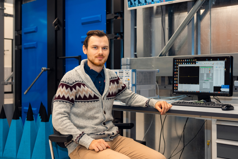
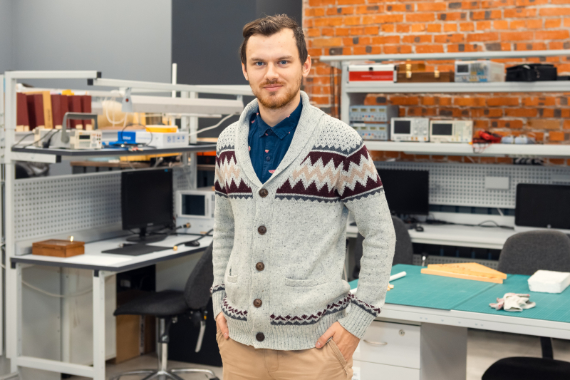
Evgeny Koreshin spoke to ITMO.NEWS about the ways to improve the existing MRI methods, the reasons we need special coils for urology purposes, as well as his inventions and participation in the forum in a blended format.
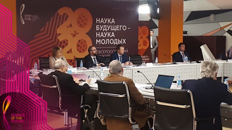
Tell us about the conference
I participated remotely. Thankfully, in the times of the pandemic, scientists came up with an idea of conducting online events (laughs). At the conference, you could quickly switch between the event’s sections, as well take part in the so-called communication rooms. But still, meeting people in person is a lot more interesting.
42 people participated in my section, all Master’s and PhD students, all with serious reports. The competition was very tough. I’m not very proficient in medicine and pharmacology, and I was quite anxious at first, but then they chose me as the winner.
The presentation contest had two rounds, the first being about presenting your work in three minutes. I wrote my speech beforehand and practiced a lot.
Why do you think your work was chosen?
Following the advice of my research supervisor, I started my presentation with the description of our research team’s general focus: coils for doing MRI of arms, resonators for other body parts, neural networks and microscopy. What’s more, my topic is well-explored, which contributed to the project being comprehensible to everyone. I did my best to use simple language, and I think that gave a good result. The head of our category, professor Boris Zhivotovsky, told me some really kind words. I was startled.
What was your presentation about?
I addressed the topic of MRI, which I focused my Master’s studies on. It's the topic of my thesis, several articles and patents. We developed a special coil for urology purposes, which was the project that I submitted for the conference.
I develop devices for MRI and the creation of anatomic images. For example, with an X-ray you can see dense tissues such as bones, and with an MRI scan — soft tissues such as muscles and fats; you can even use it to see blood vessels.
You need special devices for every part of the body, so the range of our research is quite extensive. A very good example is our project of a specialized wireless coil for diagnosing breast cancer developed by my colleagues.
My research supervisor once suggested that I develop an MRI coil for urology purposes. Men have specific genital conditions, a problem that’s solved by doctors. But there are no specialized devices they can use. And it’s us who develop such devices.
What are the benefits of your inventions?
I know that ultrasound scanning methods are used for diagnoses, but their efficiency largely depends on a doctor’s skill. MRI is a better method which makes it possible to attain better precision in noninvasive diagnosis, assess the functionality of tissue, and conduct other informative diagnosing procedures. With the help of MRI, one can get more information.
We create small coils. The smaller they are, the better, as it affects image quality. The coil has to be as close to the region of interest as possible. For example, a coil for the head goes around it, a coil for the knee is fixed tightly to the leg. This provides for quality imaging with low noise levels.
There are no coils specifically designed for urology purposes. The standard examination procedure is when the patient lies on his back, his penis is fixed on his belly, and the coil is placed on top of it. This method is associated with the organ bending and moving which has a negative effect on examination results.
We came up with the idea to have the patient lie on his belly on a special platform which offers direct access to the penis so that there are none of the aforementioned deficiencies.
What’s more, MRI can heat the patient, which has to be avoided. We conducted computer modeling, as well as experiments on a cactus in order to prove our method’s safety. Once we were sure that nothing endangers our patients, we started experimenting in-vivo.
What else does one need for a successful MRI?
The procedure has to do with placing a person in a strong magnetic field. The antenna in the tomograph transmits the radio signal to the tissue, and then the receiving antenna registers the response. Another coil is usually used for this purpose; we use a wireless resonator coil.
Our contribution is small, but very significant, as it’s important for the final stage where you get the images.
What’s the resonator for?
The resonators that we develop can be used by people with metal prostheses. Those who have them can’t make use of MRI, as metal elements can overheat due to the concentration of a high electric field. When we use a resonator, we concentrate all the energy in a small area while leaving the other parts of the body unaffected. This means that we increase the procedure’s safety, and it can now be used by those who couldn’t get MRI scanned before, and not only for urology purposes.
When a person lies in a tomograph without a resonator, we receive useful signals but also a lot of noise; the signal is received from the region of interest, and noise - from the whole body. When the process takes place in a small area, the resulting image has better quality.
What are the difficulties associated with bringing your invention to the market?
Commercial coils are easier to handle, while our device is a bit more complex and calls for adjustment. But it’s nothing impossible, we already see the steps we need to take. For example, we can improve the quality of images in the coil, as it’s still not optimal. And for now, our main goal is to make the coil simpler so that doctors will be able to use it.
And they will be ready for use at hospitals?
Yes. If you approach a radiotherapist and ask them about the use of MRI in urology, they will mention such conditions as prostate cancer or urinary tract disorders (these are the areas inside the body). The penis hasn’t been studied much with regard to the MRI methods, there are only about 10 serious research articles on this topic.
There are some specific conditions that can be seen using MRI. If you can timely identify problems with the nerve tissue, you can start treating conditions such as erectile dysfunction in advance. Another example of a disease you’ll be able to identify is the Peyronie disease that can result in penis deformation.
How will winning this contest help your career?
Our work is supported by grants, and the acknowledgement of your results by the scientific community contributes to your application being chosen. Winning at a conference is a high mark, it increases your chances at the successful development of your project.