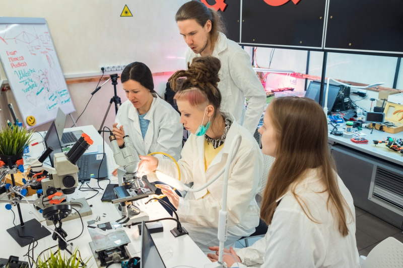
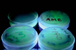
Students Maria Moschenskaya, Veronika Prizova, Galina Alferova, Evgeny Khlopotov and Siraj Farhanulla began their project Plant Vision back in November 2020. They are mentored by Ippolit Markelov, a famous artist, biologist, and founder of the 18 apples science art group. As part of a week-long workshop, the students conducted their experiment according to all research procedures, and also went on to present it at the recent Congress of Young Scientists.
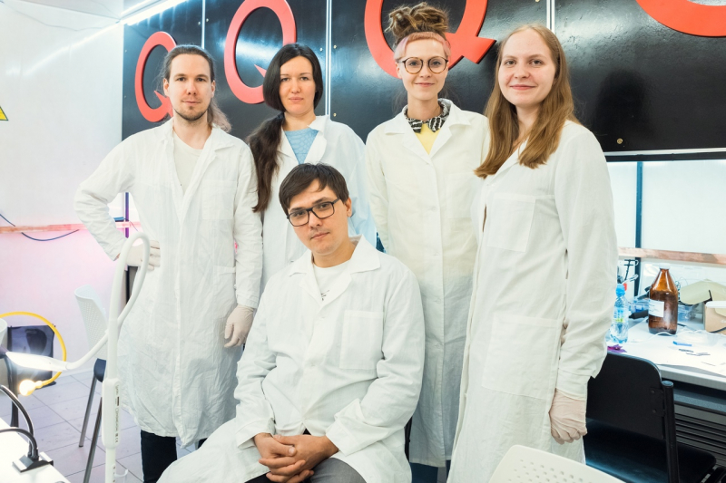
The workshop's participants: Evgeny Khlopotov, Galina Alferova, Maria Moschenskaya, Veronika Prizova, and Ippolit Markelov
The team got inspiration for their project from the recent discovery of a new species of liana native to Chile – Boquila trifoliolata. This plant that was discovered only five years ago impressed the scientists with its remarkable capacity for mimicry: it can imitate not just the color of its host plant but also its form and defects. It is still not clear how this works, how the liana collects and processes the information on its host’s looks. One of the hypotheses implies that the plant can actually “see”.
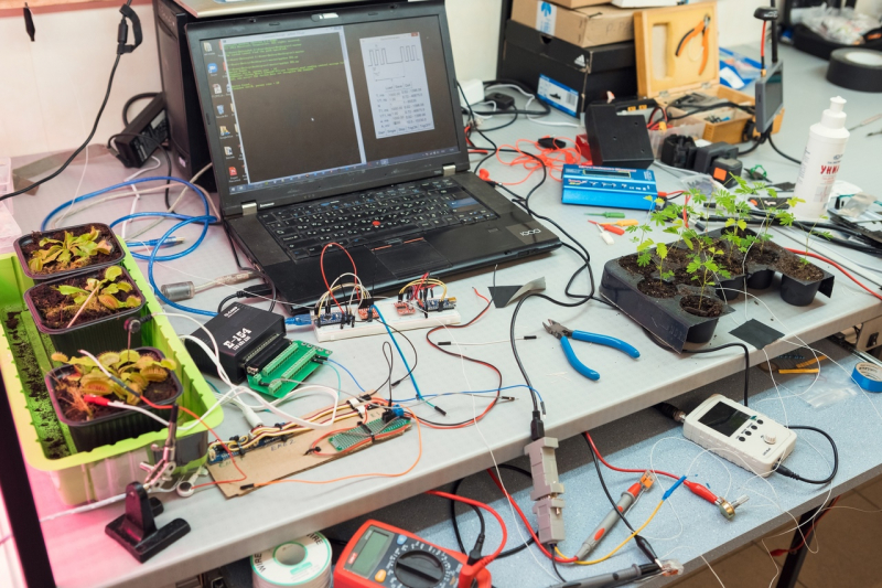
The workshop on photoreception in higher plants
Converging lens
The team took an article by Stefano Mancuso that was published in Trends in Plant Science (this journal is part of Cell Press) as their inspiration for the project. The author brings up the issue of whether plants have ocelli and refers to experiments conducted in the early 20th century by Harold Wager, Francis Darwin, and Gottlieb Haberlandt. For one, Harold Wager published a series of monochrome photographs that confirm the hypothesis that the upper layer of epidermis of higher plants acts (or can act) as a converging lens.
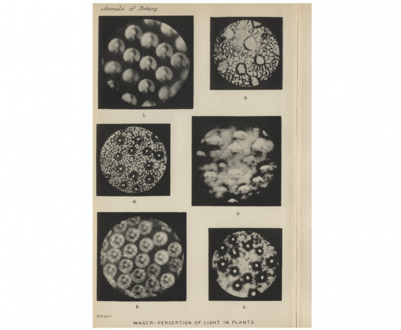
Illustration from the article by Stefano Mancuso. Credit: JSTOR Collection (Annals of Botany, 1909), Oxford University Press
“Stefan Mancuso entertains the hypothesis that higher plants have a set of organs that are essential for photoreception. This is a converging lens (the upper layer of epidermis), and photosensitive cells that act as a retina (in the mesophyll layer, under this lens). All of this suggests that higher plants have some organ of vision that – theoretically – can bring an image into focus and process it in some way,” explains Ippolit Markelov.
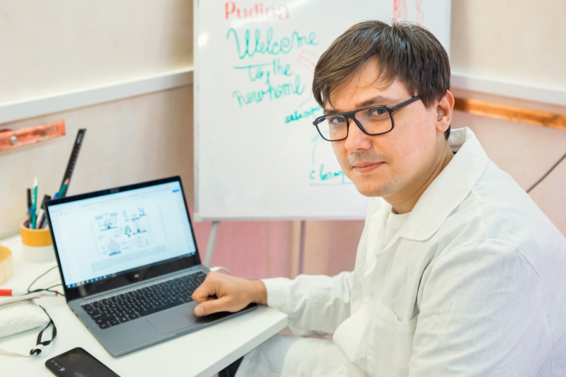
Ippolit Markelov
As a result, the team was able to repeat the 1909 experiment, and even improve it – for example, their photos have color and have a better resolution. What’s more, the students did their experiment without several components:
“We understood that we don’t need both the specimen and the cover glass. This way, the reflection is better, as there are fewer flares and diffractions. Plus, we didn’t use water and glycerin to moisten the sample – and the image turned out every bit as good as in the initial experiment. This is yet another argument in favor of the upper layer of epidermis acting as a converging lens, that the image is not only a reflection in a drop of liquid,” comments Veronika Prizova.
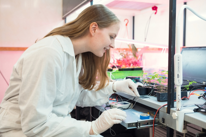
Veronika Prizova
They got the image with the help of a microscope with an 8x lens that can display in real-time mode. The sample was a monolayer of epidermis, which is essentially a layer of double convex lens, as the cells are filled with water. An image that’s prepared in advance is projected on these lenses.
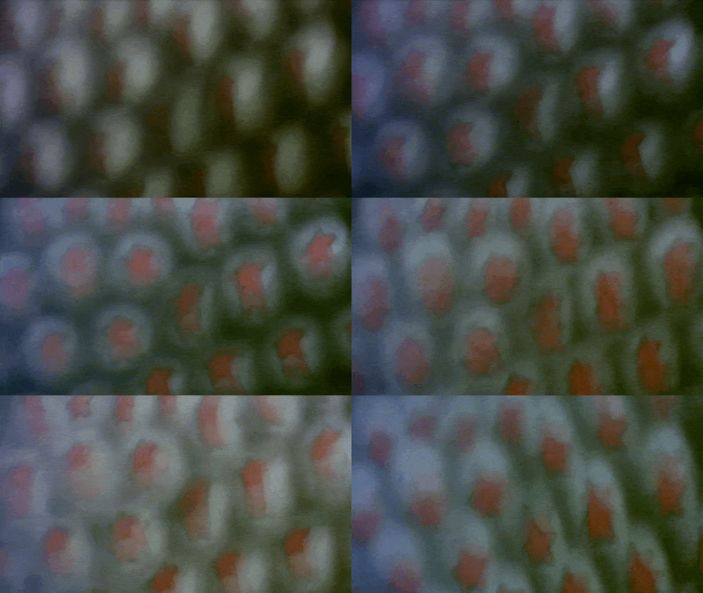
The image obtained with the help of a microscope with an 8x lens
How the plant reacts
The next stage of Plant Vision will have to do with research on the mechanism of plants’ reaction to external stimuli in general. They chose Dionaea muscipula, commonly known as Venus flytrap, as a study sample – one of the most famous and well-studied species of carnivorous plants.
“Inside the trap of Dionaea muscipula, there are six sensitive hairs – mechanoreceptors. The victim has to touch any of them twice in a row in order for the trap to react. Every time these hairs are irritated, the polarity of potentials in the membrane changes, and thanks to that the action potential accumulates. When the victim touches the hair for the second time, liquid is instantly transferred from one layer to another – this, in turn, induces a change in the leaf’s geometry and the response of the trap. If the victim is inside the trap, starts panicking and tries to get out, it activates the mechanoreceptors for the third time – and this induces the digestive secretion,” says Evgeny Khlopotov.
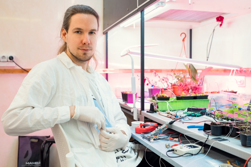
Evgeny Khlopotov
Taking the Venus flytrap as an example, one can not just study the whole complex of biophysical reactions of a plant, but also record these processes with the help of special sensors – and then imitate them with the help of electric stimulations.
“In their work, the students have the task to, first of all, study the electrochemical properties of the whole biological process, secondly, to figure out all of the current-voltage and temporal properties and, thirdly, model them with the help of electrical simulations that would imitate the operation of the trap without mechanoreceptors. Yet another task is to use it all to create an interspecies communication device (that would connect a Venus flytrap with a mimosa) that would make it possible for the plants to exchange electrical impulses between each other,” comments Ippolit Markelov.
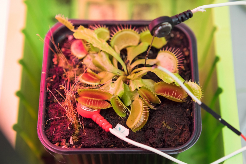
The Venus flytrap (Dionaea muscipula)
Human-plant communication
Plant Vision’s global goal is to create an artistic installation that would make it possible to establish contact between a human being and a plant. For this purpose, they will create a neural network that would translate the biochemical and biophysiological reactions inside the plant to a language that a human can comprehend.
“We will gather data: electrical signals, chemical and acoustic (ultrasound) in order to find patterns in plants’ reactions in response to external stimuli – in order to analyze them with the help of machine learning methods. As a result, the neural network will be trained to “see” what a plant sees, and process this information in a format a human can understand. This way, we will get closer to knowing how exactly plants perceive their surroundings and how they react to it,” explains Evgeny Khlopotov.
The first results were already presented at the Congress of Young Scientists at ITMO. The project will also be presented at the final exhibition of Art & Science Master’s students that will take place in late June.
